How to Isolate Exosome?
Exosomes are membrane-secreted extracellular vesicles carrying nucleic acids, proteins, lipids, and metabolites that transfer cell-specific components to recipient cells and play a critical role in intercellular communication and signaling. In recent years, the role of exosomes in pathobiological processes has attracted much attention. It has been reported that exosomes have great potential as drug carriers, as biomarkers, and for developing therapeutic approaches.
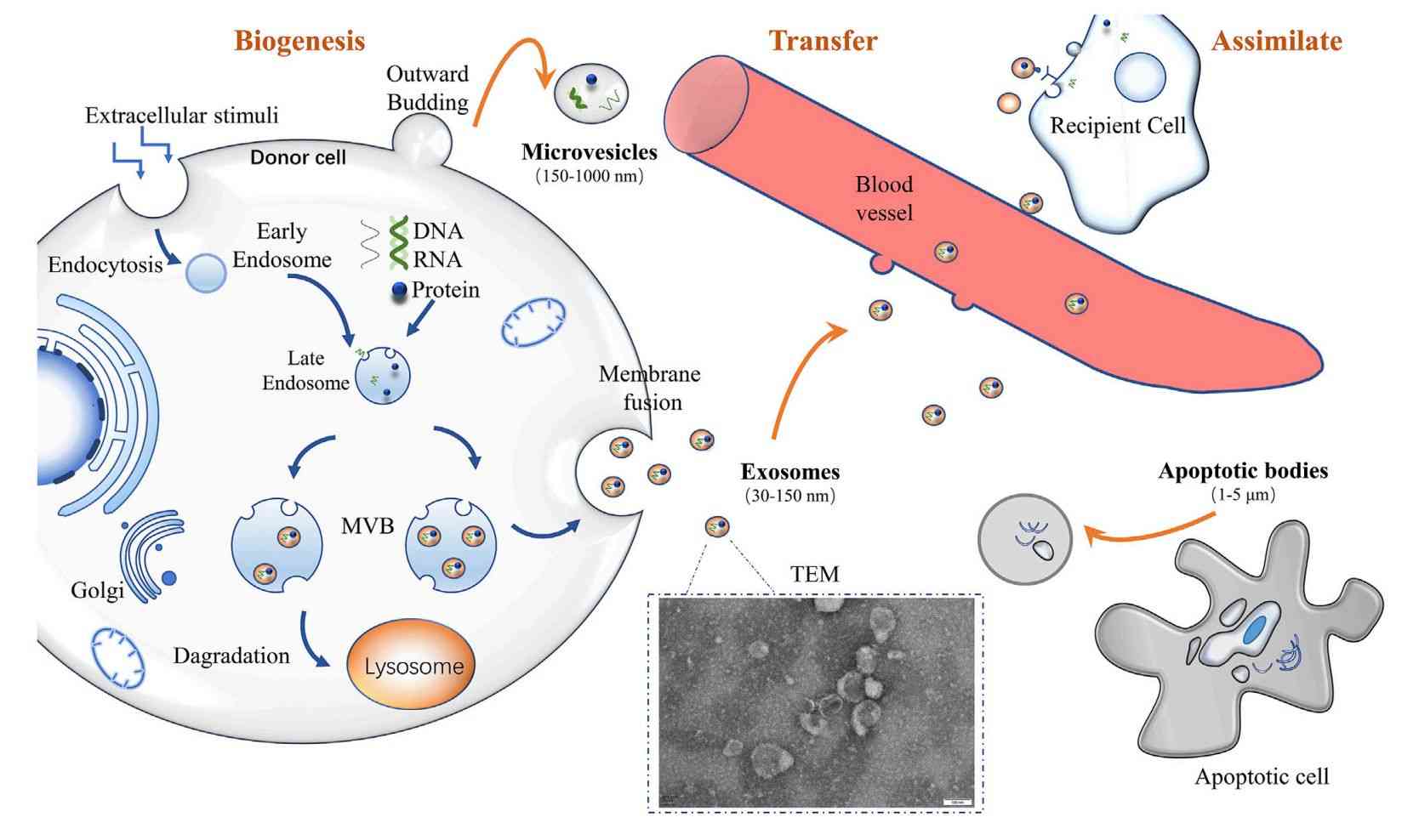
 Figure 1. Biogenesis of exosomes and other vesicles. (Chen J, et al., 2022)
What are Exosomes?
Exosomes are extracellular vesicles released by virtually all cell types and range in size from 30nm to 150nm. In addition to performing many biological functions (intercellular communication and signaling), several biological entities (e.g., proteins and microRNAs) in exosomes are closely associated with the pathogenesis of most human malignancies. Therefore, they may serve as valuable biomarkers for disease diagnosis, prognosis, and treatment.
With a great deal of research into the biological origin, material composition, and distribution in body fluids it has been found that exosomes have multiple functions. Research has found that exosomes play a critical role in the following processes:
- Immune response
- Antigen presentation
- Cell migration
- Cell differentiation
- Tumor metastasis and progression
- Apoptosis
- Angiogenesis
- Blood coagulation
Based on the great promise of exosomes as a tool for disease diagnosis and management, it is essential to isolate exosomes from various biological matrices efficiently, simply, and economically. Creative Biostructure has decades of experience in exosome research, and we have cutting-edge exosome isolation technology to provide clients with customized solutions for exosome preparation.
Our Exosome Isolation Technologies
At Creative Biostructure, we have developed and optimized methods and tools for rapid, scalable, and reproducible exosome isolation, providing high-throughput solutions for exosome heterogeneity and biomarker discovery.
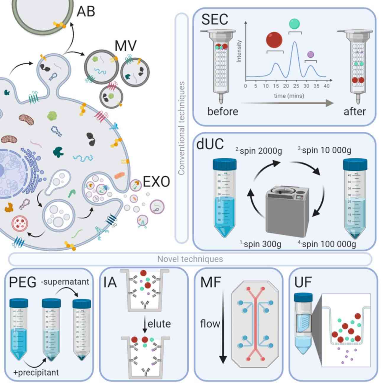
 Figure 2. Exosome isolation methods. (Sidhom K, et al., 2020)
Below is a summary of our exosome isolation methods.
| Methods | Principle | Advantages | Limitations |
| Differential Ultracentrifugation (dUC) | Using high-speed centrifugation, exosomes are separated according to their size. | Methods have been established and are widely used to purify exosomes from different biological fluids. | Laborious, time-consuming, may co-precipitate non-exosomal material and may cause damage to exosomes due to high forces. |
| Size-exclusion Chromatography (SEC) | Exosomes are separated according to size by a porous column. | Gentle method to maintain exosome integrity. | May co-elute with proteins, which requires specialized equipment. |
| Polymer Precipitation | Polymer is added to induce exosome aggregation and precipitation. | The process is simple and fast with good yields of various biofluids. | Polymer contamination and exosome integrity may be compromised. |
| Density Gradient Centrifugation (DGC) | Using gradient centrifugation, exosomes are separated based on their buoyant density. | High purity of exosomes obtained. | Complex, requiring careful gradient preparation and long procedures. |
| Immunoaffinity (IA) | Exosomes are captured based on specific surface markers. | Highly specific and capable of targeting and isolating exosome subpopulations. | Limited by availability of well-characterized antibodies; potential loss of non-targeted exosomes. |
| Microfluidics (MF) | Exosomes are separated based on size, surface markers, or acoustic properties. | Precise control, and automation potential for rare subpopulations of exosomes. | The complex operation requires technical expertise. |
| Ultrafiltration (UF) | Separation of exosomes from other particles by filtration through a membrane with a specific pore size. | Simple and relatively fast for large sample sizes. | This method may result in the co-separation of proteins and contaminants. |
What are the Applications of Exosomes?
- Diagnostic potential of exosomes
Exosomes are present in all biological fluids, based on the specificity and stability of their contents, making them useful as minimally invasive liquid biopsy tools.
- Therapeutic potential of exosomes
Exosomes are being actively explored as therapeutic agents either by themselves or as carriers for drug payload delivery. The engineering of exosomes further enhances their potential to intervene in a wide range of diseases.
- Exosome-based vaccine development
Exosomes derived from innate immune cells and tumor cells have the potential to be used as cancer vaccines. The use of exosomes to transfer membrane proteins or their contents to alter cancer cell phenotypes facilitates exosome-based vaccine development.
- Role of exosomes in regenerative medicine
Exosomes from mesenchymal stem cells are frequently used in regenerative and aesthetic medicine and can be utilized as a novel cell-free therapy for the treatment of skin diseases.
Order Our Exosome Products
In addition to exosome isolation services, Creative Biostructure also offers high-quality exosome kits. Standardized exosome isolation kits address the heterogeneity of exosomes and provide a reproducible, simple, and straightforward method of exosome preparation. Our kits make it easier for clients to isolate exosomes in the lab with comparable or better results than ultracentrifugation and without the need for specialized equipment.
| Cat No. | Product Name | |
| Exo-UEK01 | Urine Exosome Kit | Inquiry |
| Explore All Exosome Kits | ||
Furthermore, our scientists have successfully developed exosome products from different sources that have multiple applications in exosome-based research, reflecting the functional status of their parental cells. Our products include exosomes isolated from cancer cell lines, exosomes isolated from stem cell lines, exosomes isolated from immune-related cell lines, exosomes isolated from general cell lines, exosomes isolated from body fluids, fluorescent exosomes/microvesicles, lyophilized microvesicles, exosomes isolated from human disease-state body fluids, and exosomes isolated from plant. Using our exosome products as a starting point for research helps clients contribute to the advancement of science and medicine.
| Cat No. | Product Name | Source |
| Exo-CH21 | HQExo™ Exosome-A375 | Exosome derived from human malignant melanoma cell line (A375 cell line) |
| Exo-CH02 | HQExo™ Exosome-A549 | Exosome derived from human non-small cell lung cancer cell line (A549 cell line) |
| Exo-SC03 | HQExo™ Exosome-hTERT | Exosome derived from hTERT-immortalized Mesenchymal Stem Cell |
| Exo-SC04 | HQExo™ Exosome-MSC | Exosome derived from Xeno-Free Human Mesenchymal Stem/Stromal Cells and Media |
| Exo-IC01 | HQExo™ Exosome-BC3 | Exosome derived from human B lymphocyte cell line (BC-3 ) |
| Exo-IC05 | HQExo™ Exosome-JAWSII | Exosome derived from mouse bone marrow immature dendritic cell line (JAWSII) |
| Exo-GC07 | HQExo™ Exosome-CD63-EGFP | Exosome derived from human embryonic kidney cell line (HEK293, CD63-EGFP) |
| Exo-GC10 | HQExo™ Exosome-CD63-mCherry | Exosome derived from human embryonic kidney cell line (HEK293, CD63-mCherry) |
| Exo-EV-A-001 | HQExo™ Exosome-Bovine Plasma exosome | Exosome derived from Bovine Plasma |
| Exo-EV-A-002 | HQExo™ Exosome-Canine Beagle Plasma exosome | Exosome derived from Canine Beagle Plasma |
| LEV-F665-LnCAP | HQExo™ Microvesicles-665-LnCAP | Fluorescent labeled microvesicles derived from human prostate adenocarcinoma (LnCAP cell line) |
| Exo-F488-A549 | HQExo™ Exosome-488-A549 | Fluorescent labeled exosome derived from human non-small cell lung cancer cell line (A549 cell line) |
| LEV-11 | HQExo™ Microvesicles-A549 | Microvesicles derived from human non-small cell lung cancer cell line (A549 cell line) |
| LEV-13 | HQExo™ Microvesicles-B16F10 | Microvesicles derived from murine melanoma cell line (B16F10 cell line) |
| Exo-HDBF-01 | HQExo™ Exosome-SDH-Alzheimer's plasma | Exosome derived from Single Donor Human Alzheimer's plasma |
| Exo-HDBF-02 | HQExo™ Exosome-SDH-Asthma plasma | Exosome derived from Single Donor Human Asthma plasma |
| Exo-PDELN01 | HQExo™ Exosome-Garlic | Exosome derived from Garlic |
| Exo-PDELN02 | HQExo™ Exosome-Ginger | Exosome derived from Ginger |
| Explore All Exosome Products | ||
Creative Biostructure has been following advances in exosome research for many years. With expertise in exosome isolation, characterization, analysis, engineering, and application development, we provide strong support to our clients. If you are looking for high-quality exosome products and reliable exosome isolation services, welcome to contact us anytime.
References
- Chen J, et al. Review on Strategies and Technologies for Exosome Isolation and Purification. Front Bioeng Biotechnol. 2022. 9: 811971.
- Sidhom K, et al. A Review of Exosomal Isolation Methods: Is Size Exclusion Chromatography the Best Option? International Journal of Molecular Sciences. 2020. 21(18): 6466.
- Gao J, et al. Recent developments in isolating methods for exosomes. Front Bioeng Biotechnol. 2023. 10: 1100892.
- Li P, et al. Progress in Exosome Isolation Techniques. Theranostics. 2017. 7(3): 789-804.
