Structural Research of Polyomaviridae
The Polyomaviridae are a group of small, unenveloped, double-stranded DNA genome-containing viruses comprising four genera that infect mammals and birds. Typical members of the family are the JC virus (JCV) and the BK virus (BKV), which can cause significant harm and have been associated with various diseases in humans. For instance, JCV is known to cause progressive multifocal leukoencephalopathy (PML), a severe neurological disorder primarily affecting immunocompromised individuals. BKV is linked to complications in organ transplant recipients, such as nephropathy and hemorrhagic cystitis. Additionally, some polyomaviruses have been implicated in the development of certain types of cancer, including Merkel cell carcinoma. Therefore, the development of targeted antiviral therapies and prevention strategies is particularly essential.
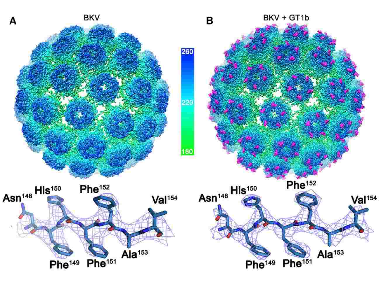
 Figure 1. The Structures of BK polyomavirus and BK polyomavirus: GT1b. (Hurdiss DL, et al, 2018)
Progress in structural research of the Polyomaviridae
In recent years, researchers have made significant progress in the structural resolution of the polyomaviruses, enhancing the understanding of the molecular structure and functional characteristics of the viruses in the family, thereby providing essential insights into their pathogenesis and potential therapeutic targets. Using cryo-electron microscopy (cryo-EM) and X-ray crystallography have determined the high-resolution structures of various polyomaviruses, visualizing the intricate details of viral capsids, viral proteins, and their interactions with host factors. For example, the structures of the major capsid proteins (VP1, VP2, and VP3 proteins) have been elucidated, providing insight into the overall organization, antigenic sites, and assembly processes. In addition, researchers have identified the large T antigen (LTAg), which plays an essential role in viral DNA replication and transcription. Structural research has revealed the structural domains and functional motifs of LTAg, shedding light on its interactions with cytokines and providing potential targets for antiviral drug development.
Particle Structure of Polyomaviruses
Viral particles are typically 40-45nm, and the capsid consists of 72 capsid grains, each consisting of five molecules of the major capsid protein VP1 and one or two minor components (VP2 and VP3). The VP1 pentamer forms a closed icosahedral capsid, and inside the capsid, each pentamer is associated with a molecule of either VP2 or VP3. Researchers used high-resolution cryo-EM to determine the structure of BKV at a resolution of 3.8 Å. The BKV capsid has T = 7d quasi-symmetry, and the six different conformations of VP1 present within the asymmetric unit of the capsid all share a core fold. A highly conserved small hydrophobic helix is present in the VP2/3 sequence of polyomaviruses.
Structural research on polyomaviruses has significantly expanded the understanding of virus molecular structure and protein function. To help clients better resolve viral structures, Creative Biostructure provides high-purity virus-like particles (VLPs) products that pave the way for the development of targeted therapeutic strategies and vaccines, improved diagnostics, and a deeper understanding of the pathogenesis of diseases related to the Polyomaviridae.
| Cat No. | Product Name | Virus Name | Source | Composition |
| CBS-V660 | JC virus VLP (capsid protein VP1 Proteins) | John Cunningham virus | E. coli recombinant | Capsid protein VP1 |
| CBS-V679 | Mouse polyomavirus VLP (VP1 Protein) | Murine polyomavirus | Insect cell recombinant | VP1 |
| CBS-V680 | Murine polyomavirus VLP (VP1; VP2 Proteins) | Murine polyomavirus | Insect cell recombinant | VP1; VP2 |
| Explore All Polyomaviridae Virus-like Particle Products | ||||
Based on knowledge of viral structural biology and long-term project experience, Creative Biostructure's team of scientists has a deep understanding of the structural resolution of viral particles and VLPs. We have advanced equipment to accurately characterize the morphology of viral particles including high-resolution cryo-electron microscopy (cryo-EM), to support the development of targeted antiviral therapies and vaccines.
In addition, our custom VLP construction services can satisfy the specific requirements for formulating and modifying target VLPs. For a detailed quotation, please feel free to contact us.
References
- Hurdiss DL, et al. The Structure of an Infectious Human Polyomavirus and Its Interactions with Cellular Receptors. Structure. 2018. 26(6): 839-847. e3.
- Moens U, et al. ICTV Virus Taxonomy Profile: Polyomaviridae. J Gen Virol. 2017. 98(6): 1159-1160.