Introduction: The Importance of Crystallography
Crystallography plays a vital role in structural biology, materials science, and drug discovery by providing detailed insights into molecular structures. Through diffraction techniques, scientists can determine the precise arrangement of atoms in a crystal, which helps us understand the properties, behaviors, and interactions of various substances at the atomic level. The analysis of crystal structures is particularly crucial in drug development, where knowing the structure of a compound allows researchers to optimize its efficacy, stability, and interaction with target molecules.
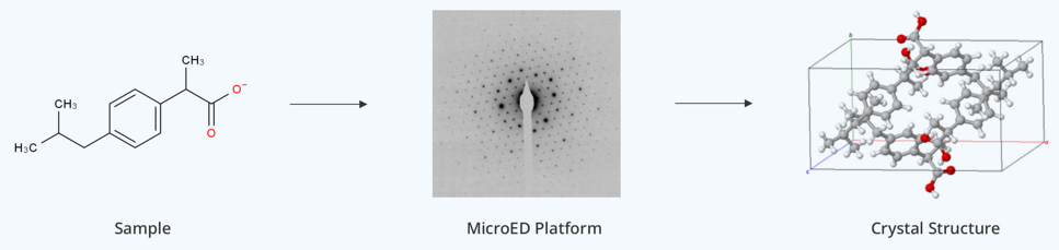
In this article, we'll explore two prominent crystallography techniques—Single Crystal X Ray Diffraction (SCXRD) and Micro-crystal Electron Diffraction (MicroED)—highlighting their principles, advantages, limitations, and applications across diverse fields. Understanding their differences will enable researchers to choose the most suitable method for their projects.
What is Single Crystal X Ray Diffraction (SCXRD) Technique?
Principle of Single Crystal XRD
SCXRD is the "gold standard" technique for determining the three-dimensional structure of molecules in single crystal form. This method involves directing X-rays at a high-quality single crystal, where the X-rays are diffracted by the atoms in the crystal lattice. The diffraction patterns obtained are analyzed to reveal the positions of atoms within the crystal, enabling precise 3D structure determination.
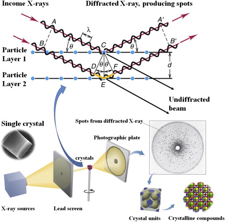 Figure 1. Schematic representation of single crystal X-ray diffraction, illustrating sample interaction with X-rays, data refinement, and structure analysis. (Liu J L, et al., 2015)
Advantages of SCXRD
- High Resolution: SCXRD offers extremely high-resolution data (up to sub-angstrom level), suitable for accurate determination of hydrogen atom positions. This technique can provide atomic-level precision, including bond lengths, angles, and intermolecular interactions.
- Widely Established: SCXRD is a well-established technique, supported by years of research and a large amount of available software for data analysis.
Limitations of SCXRD
- Dependence on High-Quality Single Crystals: SCXRD requires the cultivation of large, high-quality single crystals, typically larger than 10 μm in size. The process of obtaining these crystals can take weeks or even months, which is a significant time investment. For some materials—such as nanoparticles, small organic molecules, or certain inorganic compounds—growing suitable single crystals is often difficult or even unachievable. In addition, SCXRD is not suitable for analyzing materials with defects, nano-sized crystals, or complex structures such as co-crystals or polymorphs.
- Sample Quantity Requirements: SCXRD generally demands a substantial amount of sample material, which can be challenging, particularly in high-stakes fields like drug discovery, where sample costs can be high. Obtaining sufficient quantities of high-quality single crystals often presents a bottleneck, limiting the technique's scalability and accessibility for some research projects.
- Radiation Damage: High-energy X-rays used in SCXRD can sometimes cause radiation damage to the sample, especially in the case of sensitive materials like Metal-Organic Frameworks (MOFs). This can lead to the loss of crystallinity or the degradation of the sample during data collection, affecting the quality of the resulting structural analysis.
- Time-Consuming Process: The entire process—from crystal growth to data collection and structural analysis—can be extremely time-consuming. The need for long-term crystal cultivation, followed by detailed diffraction analysis, can delay research projects. This makes SCXRD less suited to high-throughput studies or projects where quick results are necessary.
What is Microcrystal Electron Diffraction (MicroED)?
Principle of MicroED
Micro-crystal Electron Diffraction (MicroED) is an emerging crystallographic technique that utilizes electron beams as the light source to analyze the structures of very small crystals, typically at the micrometer or even nanometer scale. The principle behind MicroED is based on the strong interaction between the electron beam and the nanocrystal. The electron beam is directed at the sample, and as it interacts with the crystal lattice, diffraction patterns are generated. These diffraction patterns are then collected, typically using continuous rotation techniques such as the Rotation Electron Diffraction (RED) or Automated Diffraction Tomography (ADT) methods. These techniques enable the collection of three-dimensional diffraction data, which is essential for determining the precise atomic-level arrangement of molecules in the crystal.
MicroED can provide similar structural information to traditional X-ray diffraction, including:
- Molecular Structure: Determining the three-dimensional atomic positions, bond lengths, bond angles, and molecular configuration.
- Crystal Packing: Analyzing how molecules are arranged and packed within the crystal lattice, including the spatial distribution of atoms.
- Polymorphism: MicroED can effectively reveal the internal structures of different polymorphs, especially in multi-crystal or mixed-phase systems.
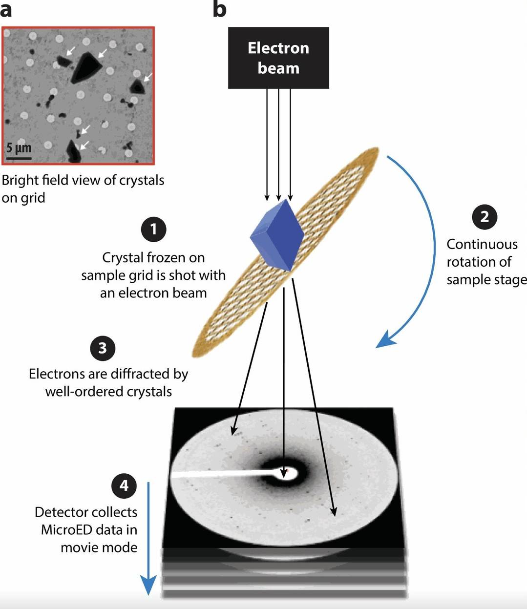 Figure 2. Overview of Microcrystal Electron Diffraction (MicroED). MicroED workflow: (a) Screening microcrystals on an electron microscope grid. (b) Diffraction data collection from rotating microcrystals in electron beam mode. (Xu Y, et al., 2021)
Advantages of MicroED
- No Need for Large Single Crystals: One of the key advantages of MicroED over SCXRD is that it doesn't require the cultivation of large, high-quality single crystals. This avoids the long and labor-intensive process of single crystal growth, significantly improving efficiency, especially for samples that are difficult to crystallize.
- Minimal Sample Requirement: MicroED requires only tiny amounts of sample, as small as 1 mg or even nano-sized crystals (100 nm). This is particularly useful for rare or difficult-to-crystallize compounds, such as natural products or small biomolecules. For example, MicroED has been used to analyze nanocrystals of cytochrome P450.
- Flexible Sample Adaptability: Unlike SCXRD, MicroED can handle polycrystalline, mixed-crystal, and disordered systems. It can also analyze compounds that are hard to crystallize, offering versatility across a wide range of materials. In some cases, it even allows for direct determination of hydrogen atom positions.
- Efficient Data Collection: Using rotational techniques like RED or ADT, MicroED can collect data quickly—typically within a few hours. This speed makes it an excellent choice for high-throughput analysis, allowing researchers to process multiple samples in a short time frame.
- Direct Mapping of Protons and Hydrogen Bonds: Since MicroED uses electron beams, it can more accurately map electron density, allowing for the direct determination of proton positions and hydrogen bond networks. This capability enriches the structural information and is particularly beneficial for studying complex biomolecules where hydrogen bonding plays a key role.
Limitations of MicroED
- Resolution: While MicroED provides high-quality structural data, it generally offers slightly lower resolution than SCXRD, especially for complex structures.
- Dynamical Scattering Effects: MicroED can be affected by dynamical scattering, where the diffraction intensity may be skewed due to multiple scattering events. This requires the use of specialized algorithms for intensity correction, adding complexity to data analysis.
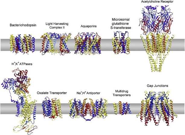
- Electron Beam-Induced Damage: The high-energy electron beams used in MicroED can cause damage to the sample, especially for sensitive materials. For example, when studying membrane proteins embedded in lipids, rapid freezing is often necessary to minimize radiation-induced damage.
SCXRD vs MicroED: Key Differences in Sample Requirements
SCXRD Sample Requirements
- Sample Size and Quality: SCXRD requires high-quality single crystals that are large enough to diffract X-rays effectively. Typically, these crystals must be at least 0.3 mm in size and must be free of cracks or defects. Additionally, the crystal must be transparent to X-rays to produce high-quality data.
- Best Use: Best suited for samples that can form high-quality single crystals, including small organic molecules, inorganic crystals, and well-defined biomolecules.
MicroED Sample Requirements
- Minimal Sample Requirement: MicroED requires significantly less material. It can analyze crystals as small as 100 nm to a few microns in size. This makes MicroED ideal for analyzing samples that are difficult or impossible to grow as large single crystals, such as nanomaterials, complex organic molecules, or proteins embedded in membranes.
- Best Use: It is well-suited for small amounts of rare compounds or when sample preparation is difficult. MicroED can also handle polycrystalline and mixed-crystal samples, providing flexibility in dealing with complex sample systems.
Comparison Table: Sample Requirements
| Criteria | SCXRD | MicroED |
|---|---|---|
| Sample Size | ≥0.3 mm (large, high-quality single crystals) | 100 nm to several microns (nano and micro crystals) |
| Crystal Quality | Requires transparent, crack-free single crystals | Requires small crystals, less focus on size/quality |
| Sample Quantity | Requires significant sample quantity | Minimal sample requirement (1 mg or less) |
| Best Use | Best for samples with good crystallization capabilities | Best for nanocrystals, membrane proteins, and rare materials |
SCXRD vs MicroED: Key Differences in Light Source and Instrumentation
SCXRD Light Source and Instrumentation
- Light Source: SCXRD uses X-rays as the light source. These can be generated using laboratory X-ray systems or synchrotron radiation sources.
- Instrument Requirements: SCXRD typically requires either an X-ray generator or access to synchrotron facilities. Synchrotrons provide stronger X-ray beams, making them necessary for weakly diffracting samples. Laboratory X-ray systems work well for routine analyses but may struggle with weak samples.
- Best Use: Best suited for large, well-formed crystals that can be easily placed in an X-ray beam. Commonly used for routine analyses of high-quality samples in laboratory settings.
MicroED Light Source and Instrumentation
- Light Source: MicroED uses an electron beam, which has a much shorter wavelength than X-rays. This allows the electron beam to interact strongly with even the smallest crystals, producing high-quality diffraction patterns.
- Instrument Requirements: MicroED requires a cryo-transmission electron microscope (cryo-TEM), which operates at cryogenic temperatures to preserve the sample, especially for sensitive materials like proteins or lipids.
- Best Use: Ideal for small, weakly scattering, or radiation-sensitive samples, such as nanocrystals, organic compounds, or biological molecules like membrane proteins.
Comparison Table: Light Source and Instrumentation
| Criteria | SCXRD | MicroED |
|---|---|---|
| Light Source | X-rays (lab or synchrotron) | Electron beam (shorter wavelength) |
| Instrument | X-ray diffractometer, synchrotron source | Cryo-TEM |
| Challenges | Requires synchrotron for weak samples | Requires cryogenic conditions, complex setup |
| Best Use | High-quality single crystal analysis | Small, weakly scattering, or sensitive samples |
SCXRD vs MicroED: Key Differences in Data Analysis
SCXRD Data Analysis
- Resolution and Precision: SCXRD provides very high-resolution data, typically achieving sub-angstrom precision. It is excellent for accurately determining the positions of light atoms, such as hydrogen, and for defining absolute configurations of molecules.
- Data Processing: SCXRD data processing is well-established with software like SHELX, which allows for efficient structure determination and refinement. The process is mature and reliable.
- Best Use: SCXRD is ideal for studies that require high-resolution, precise structure determination, especially with well-formed, large crystals.
MicroED Data Analysis
- Resolution and Precision: MicroED achieves similar resolution to SCXRD, but the data can be affected by dynamical scattering, especially in complex samples. This can reduce the precision of the results in certain cases.
- Data Processing: MicroED requires specialized software like DIALS or XDS, which need more complex corrections and refinement due to the nature of electron diffraction data. The process is still developing and can be more challenging than traditional X-ray diffraction.
- Best Use: MicroED is useful for quickly analyzing small, weakly scattering, or complex samples where speed is important, but it may not offer the same precision as SCXRD for high-resolution work.
Comparison Table: Data Analysis
| Criteria | SCXRD | MicroED |
|---|---|---|
| Resolution | Sub-angstrom precision | Similar to SCXRD, but affected by scattering effects |
| Precision | Very high, especially for light atoms and configurations | Lower precision in complex systems due to scattering |
| Data Processing Software | SHELX (efficient and well-established) | DIALS, XDS (more complex and evolving) |
| Challenges | Dependent on sample quality and synchrotron access | Sensitive to sample conditions and requires advanced corrections |
| Best Use | High-resolution, detailed structure analysis | Fast analysis for small or complex samples |
SCXRD vs MicroED: Key Differences in Efficiency
SCXRD Efficiency
- Sample Preparation: SCXRD needs a significant amount of time for sample preparation. Growing large, high-quality single crystals can take days to weeks, especially for difficult samples. This step is time-intensive because high-quality, defect-free crystals are required.
- Data Collection and Processing: After the sample is ready, collecting data and processing it can take additional time, particularly for complex structures.
- Best Use: SCXRD is ideal for detailed studies of well-formed crystals, where accuracy is more important than speed.
MicroED Efficiency
- Sample Preparation: MicroED requires much less time for sample preparation. It only needs a small amount of material and the process can be completed in hours.
- Data Collection and Processing: MicroED allows for faster data collection and processing. Testing can be completed in just a few hours, making it a great choice for fast, high-throughput analysis.
- Best Use: MicroED is perfect for projects that require quick results or when working with multiple samples.
Comparison Table: Time and Efficiency
| Criteria | SCXRD | MicroED |
|---|---|---|
| Sample Preparation Time | Days to weeks (especially for hard-to-crystallize samples) | Hours, minimal material required |
| Data Collection Time | Longer (takes days for large crystals) | Shorter (takes hours for small crystals) |
| Data Processing Time | Time-consuming, especially for complex samples | Faster, with specialized software |
| Best Use | High-precision analysis of well-formed crystals | Quick results, high-throughput screening |
SCXRD vs MicroED: Key Differences in Application Areas
When considering their application scenarios, X ray diffraction from single crystal and MicroED serve distinct technical needs while also complementing each other in certain contexts.
Biological Research
- SCXRD: In structural biology, SCXRD remains the gold standard for determining high-resolution 3D structures of proteins, nucleic acids, and other biomolecules, requiring large, high-quality single crystals.
- MicroED: MicroED addresses the limitations of SCXRD when dealing with small, challenging crystals that cannot be grown large enough for single-crystal diffraction. It offers a flexible approach for studying biological macromolecules, such as proteins and nucleic acids, especially when single crystals are difficult to obtain.
Pharmaceutical Development
- SCXRD: This technique is invaluable for accurately determining the crystal structures of drug molecules, including the precise analysis of chirality centers. It is essential for detailed studies of drug stability, polymorphs, and interactions at the molecular level.
- MicroED: Unlike SCXRD, MicroED is particularly useful in the rapid screening of drug polymorphs. It also helps investigate the interaction between drugs and proteins, aiding in drug design and development by providing faster data on crystal forms and molecular interactions.
Material Science
- SCXRD: SCXRD excels in providing high-resolution structural data for high-quality crystalline materials, making it ideal for studying their microstructural properties in detail. It is used extensively in materials science for precision work involving well-formed crystals.
- MicroED: MicroED overcomes the limitation of SCXRD when studying nanomaterials, amorphous materials, and framework compounds (such as MOFs and COFs). This method is well-suited for complex structures where traditional SCXRD might struggle due to sample size and quality constraints.
Comparison Table: Application Areas
| Criteria | SCXRD | MicroED |
|---|---|---|
| Pharmaceutical Development | Precise analysis of drug crystal structures and chirality centers | Rapid polymorph screening and drug-protein interactions |
| Biological Research | High-quality single-crystal studies of proteins, nucleic acids | Analysis of microcrystals and difficult-to-grow biomolecules |
| Material Science | High-resolution structural analysis of high-quality crystals | Analysis of nanomaterials, amorphous materials, and MOFs/COFs |
| Sample Flexibility | Best for high-quality single crystals | Suitable for microcrystals, polycrystals, and complex mixtures |
| Resolution and Precision | High-resolution, ideal for detailed structural work | Comparable resolution but with the potential for scattering issues in complex systems |
In conclusion, SCXRD is particularly suited for high-resolution studies of high-quality single crystals, where precise structural data is required, such as in pharmaceuticals and material science. MicroED, with its flexibility for small and challenging samples, offers an innovative solution for rapid data collection and is ideal for microcrystals, polycrystalline systems, and amorphous materials. When combined, SCXRD and MicroED can complement each other to cover a broad range of sample types and research needs, from high-quality single crystals to complex mixtures and nanomaterials.
At Creative Biostructure, we offer both Single Crystal X-ray Diffraction (SCXRD) and MicroED services to help you achieve precise structural analysis for your research projects. Whether you're working with high-quality single crystals or challenging microcrystals, our expert team is equipped to support your needs across drug discovery, materials science, and structural biology. Contact us to discuss how our advanced crystallography solutions can accelerate your research and provide the insights you need.
References
- Sheldrick G M. Crystal structure refinement with SHELXL. Crystal Structure Communications. 2015, 71(1): 3-8.
- Liu J L, Bashir S. Advanced nanomaterials and their applications in renewable energy. 2015.
- Li J, Sun J. Application of X-ray diffraction and electron crystallography for solving complex structure problems. Accounts of Chemical Research. 2017, 50(11): 2737-2745.
- Liu S, Hattne J, Reyes F E, et al. Atomic resolution structure determination by the cryo‐EM method MicroED. Protein Science. 2017, 26(1): 8-15.
- Mu X, Gillman C, Nguyen C, et al. An overview of microcrystal electron diffraction (MicroED). Annual Review of Biochemistry. 2021, 90(1): 431-450.
- Danelius E, Halaby S, van der Donk W A, et al. MicroED in natural product and small molecule research. Natural Product Reports. 2021, 38(3): 423-431.
- Martynowycz M W, Shiriaeva A, Ge X, et al. MicroED structure of the human adenosine receptor determined from a single nanocrystal in LCP. Proceedings of the National Academy of Sciences. 2021, 118(36): e2106041118.
- Yokoo H, Aoyama Y, Matsumoto T, et al. Rapid Structure Determination of Ranitidine Hydrochloride API in Two Crystal Forms Using Microcrystal Electron Diffraction. Chemical and Pharmaceutical Bulletin. 2024, 72(5): 471-474.

-1.jpg)