MALDI-MSI Service
Matrix-assisted laser desorption ionization mass spectrometry imaging (MALDI-MSI) stands as a transformative technique in molecular imaging and biomedical research, offering unprecedented insights into the molecular architecture of tissues and cells. Creative Biostructure's MALDI-MSI service enables researchers to gain molecular insights efficiently and accurately, supporting projects from basic research to preclinical studies.
Why Perform MALDI-MSI?
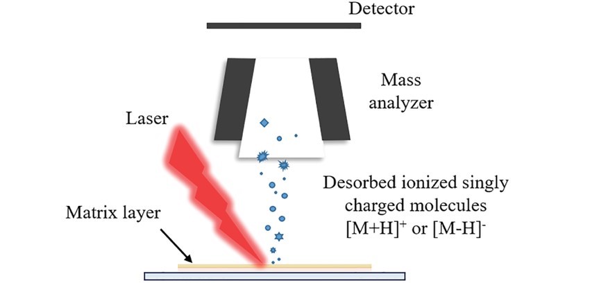
As a powerful analytical method, MALDI provides spatial resolution and molecular specificity, facilitating label-free tracking of endogenous and exogenous compounds. Similar to immunohistochemistry, MSI provides high throughput scanning of compounds, but does not require antibodies. During sample preparation for MALDI-MSI, the target tissue is sliced into thin slices and embedded in an organic matrix. The matrix promotes the desorption and ionization of the target compound under the action of an ultraviolet laser beam, and the mass-to-charge ratio of the generated ions is measured by the mass spectrometer on a programmed ablation point array, which can simultaneously identify and quantify multiple analytes. Therefore, MALDI-MSI can quickly snapshot the molecular distribution at a specific location on the tissue surface.
Since its inception, MALDI-MSI has demonstrated many unique advantages in biomedical research, which not only make it a powerful molecular imaging tool, but also promote its wide application in many fields.
- No labeling required: Unlike traditional immunohistochemistry or fluorescent labeling techniques, MALDI-MSI can obtain information directly from unprocessed tissue samples without any specific labeling of target molecules.
- High spatial resolution: MALDI-MSI systems are able to achieve spatial resolutions of up to 5 to 10 microns, providing very detailed molecular distribution maps.
- Non-destructive sample preparation: Compared with some techniques that require complete dissolution or fragmentation of the sample, MALDI-MSI uses gentle sample handling methods.
- Fast and efficient data acquisition: Advanced MALDI-MSI platforms can complete the data acquisition process at an extremely high speed and can obtain a large amount of high-quality data in a short time.
 Figure 1. The ionization principle for MALDI-MSI. (Hou Y, et al., 2023)
Our MALDI-MSI Service Procedure
We provide high-quality MALDI-MSI services, which can perform high-resolution spatial distribution analysis of target molecules such as small molecules, peptides and proteins in biological samples.
- Initial Consultation and Project Design: We start by consulting with clients to clarify research goals and tailor MALDI-MSI parameters accordingly, ensuring project-specific technical optimization.
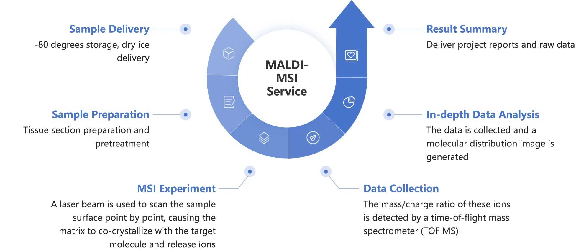
- Sample Delivery: Samples are stored at -80°C and shipped on dry ice to preserve molecular integrity and prevent degradation before analysis.
- Sample Preparation: We meticulously prepare tissue samples, including sectioning and pretreatment, to enhance molecular mapping accuracy for MALDI-MSI.
- MSI Experiment: In the MALDI-MSI process, a finely tuned laser beam systematically scans the tissue sample surface point by point. During this scanning, the applied organic matrix co-crystallizes with target molecules, releasing ions that are critical for mass spectrometric analysis.
- Data Collection: As ions are generated, they are analyzed by a time-of-flight mass spectrometer (TOF MS), which detects their mass-to-charge ratios. This step is essential for obtaining detailed molecular profiles, capturing high-resolution spatial and molecular specificity.
- In-depth Data Analysis: We analyze the data to produce molecular distribution images, offering insights into biomolecule localization and tissue composition.
- Result Summary: We provide a comprehensive report with raw data and customized images, supporting client research objectives and facilitating further study or data integration.
 Figure 2. Our MALDI-MSI service workflow. (Creative Biostructure)
Why Choose Creative Biostructure?
- Expert Support: Led by seasoned scientists, our team provides end-to-end guidance, ensuring top-tier scientific rigor and optimized outcomes in MALDI-MSI and related projects.
- State-of-the-Art Technology: We employ high-resolution, swift imaging systems for superior spatial detail and rapid data collection, enabling precise molecular mapping suitable for complex research demands.
- Tailored Services: We customize our approach to fit each project's unique requirements, working closely with clients to fine-tune experimental parameters to meet their research goals.
- Rapid Turnaround: Our streamlined processes ensure swift data analysis and reporting, providing researchers with comprehensive insights and results in a timely manner.
Frequently Asked Questions
-
What types of samples can be processed by your MALDI-MSI service?
Our MALDI-MSI service can process a variety of biological samples, including but not limited to fresh frozen tissue sections, plant tissues, and insect samples. We can also image and analyze different types of molecules such as proteins, peptides, lipids, metabolites, and drugs.
-
What does your data analysis service include?
Our data analysis service includes data preprocessing, image generation, molecular distribution mapping, and detailed statistical analysis. We also provide customized data analysis reports to help you interpret the experimental results and provide professional scientific advice based on your research needs.
-
What is the typical turnaround time for MALDI-MSI service?
Project size and complexity determine our turnaround times, yet our optimized processes ensure swift data collection and analysis.
Creative Biostructure specializes in delivering exceptional MALDI-MSI services, leveraging our expertise to cater to the broad spectrum of our clients' requirements. Should you be intrigued by our offerings, please contact us to discuss and obtain a comprehensive cost estimate.
Ordering Process
References
- Hou Y, Gao Y, Guo S, et al. Applications of spatially resolved omics in the field of endocrine tumors. Frontiers in Endocrinology. 2023. 13: 993081.
- Kajimura M, Nakanishi T, Takenouchi T, et al. Gas biology: tiny molecules controlling metabolic systems. Respiratory Physiology & Neurobiology. 2012. 184(2): 139-148.
- Chen K, Baluya D, Tosun M, et al. Imaging mass spectrometry: a new tool to assess molecular underpinnings of neurodegeneration. Metabolites. 2019. 9(7): 135.
