Liposome Spatial Omics Service
In biological systems, the spatial organization of cells and molecules is fundamental to understanding tissue function and disease mechanisms. Spatial omics technology represents a revolutionary advancement that bridges the gap between molecular profiling and spatial context, particularly crucial for liposome-based drug delivery systems. Creative Biostructure's professional team uses advanced spatial omics technology to provide a full range of services including liposome labeling, distribution visualization and drug delivery efficiency evaluation to ensure that customers obtain accurate research results.
Why Spatial Analysis Matters for Liposome Research
Liposomes, self-assembled from phospholipids, form closed spherical structures with monolayer or multilayer membranes resembling cell membranes. They offer biocompatibility, degradability, and protect encapsulated drugs from enzymatic degradation, enhancing stability and reducing toxicity. Surface modifications allow for targeted delivery and prolonged retention at lesion sites, making liposomes efficient drug carriers. Cationic liposomes, in particular, are adept at encapsulating and delivering nucleic acids like DNA and RNA.
Spatial omics technology enables the visualization of liposomes and their drug cargo, providing detailed observations of their distribution, accumulation, and dynamics within tissues and cells. This technology elucidates the precise location of liposomes and the delivery pathway of drugs to target sites, aiding in the optimization of liposome design for enhanced drug delivery efficacy.
The effectiveness of liposome-mediated drug delivery heavily depends on their spatial distribution within tissues. Unlike conventional analytical methods, spatial omics provides precise insights into:
- Cellular uptake patterns across different tissue regions
- Drug release kinetics in specific microenvironments
- Interaction dynamics between liposomes and target cells
- Biodistribution patterns at both tissue and cellular levels
 Figure 1. Spatial omics datasets from multiple experimental techniques. (Palla G, et al., 2022)
Our Comprehensive Service Portfolio for Liposome Spatial Omics
Creative Biostructure offers state-of-the-art spatial omics analysis for liposome research, utilizing multiple cutting-edge platforms:
Advanced Imaging Technologies
- MERFISH (Multiplexed Error-Robust Fluorescence in situ Hybridization)
- Visium Spatial Gene Expression
- High-resolution confocal microscopy
- Advanced light sheet microscopy
Analytical Capabilities
- Spatial resolution: Down to 100 nm
- Detection sensitivity: Single-molecule level
- Multiplexing capacity: Up to 100 targets simultaneously
- 3D reconstruction capabilities
Our Workflow of Liposome Spatial Omics Service
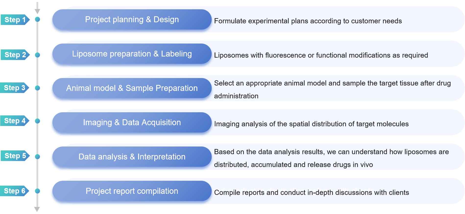
- Project Planning and Design: Based on comprehensive client consultations, we formulate detailed experimental plans according to customer needs. This includes protocol design, timeline establishment, and complete risk assessment to ensure project success. Our expert team works closely with clients to define clear objectives and deliverables, ensuring alignment with research goals.
- Liposome Preparation & Labeling: We prepare high-quality liposomes with precise size control (50-200 nm) and customize them with fluorescent labels or functional modifications as required. Each batch undergoes rigorous quality control to ensure stability and uniformity, with surface modifications tailored to specific research needs.
- Animal Model & Sample Preparation: Our team selects the most appropriate animal model based on your research objectives and administers the liposome formulation following standardized protocols. After careful timing, we collect and process target tissues using optimized preservation methods to maintain sample integrity for spatial analysis.
- Imaging & Data Acquisition: Using advanced spatial omics platforms including MERFISH and Visium technology, we perform comprehensive imaging analysis of the spatial distribution of target molecules. Our high-resolution imaging capabilities ensure accurate visualization of liposome distribution patterns across tissue sections.
- Data Analysis & Interpretation: Through sophisticated data analysis, we provide detailed insights into how liposomes are distributed, accumulated, and release drugs in vivo. Our analysis includes spatial pattern recognition, quantitative assessment of distribution, and correlation with biological parameters to ensure meaningful results.
- Project Report Compilation: We deliver comprehensive reports detailing all experimental procedures, results, and interpretations. Our team conducts in-depth discussions with clients to ensure a complete understanding of the findings and provide recommendations for future research directions. The report includes publication-ready figures and complete statistical analysis.
 Figure 2. Our liposome spatial omics service workflow. (Creative Biostructure)
Why Choose Creative Biostructure?
- Rich research experience: We have many years of experience in liposome research and development, have accumulated deep industry knowledge and technical expertise, and is able to provide customers with high-quality liposome preparation and liposome analysis services.
- Advanced technology platform: We are equipped with a series of cutting-edge technology platforms to ensure the accuracy and reliability of data.
- Professional technical team: Our team is composed of senior scientists and technical experts, providing customers with full support from experimental design to data analysis.
- Customized solutions: We provide one-stop service according to the specific needs of customers.
Frequently Asked Questions
-
How do you ensure the quality and stability of our liposomes?
We apply strict quality control standards, and we use advanced liposome characterization techniques to ensure that the particle size, morphology, and stability of our liposomes meet the highest standards.
-
How can liposomes be fluorescently labeled for visualization?
Liposomes can be labeled by incorporating fluorescent dyes or fluorescent proteins into the lipid bilayer.
-
How do you ensure the stability of fluorescent labels during analysis?
We employ photobleaching-resistant fluorophores and optimize imaging conditions to maintain signal integrity throughout the analysis.
-
Can you analyze custom-modified liposomes?
Yes, our protocols can be adapted for various liposome modifications, such as novel targeting ligands, custom fluorescent tags and specialized surface modifications.
Creative Biostructure offers high-quality liposome-related services tailored to your specific needs, leveraging our expertise in technical knowledge. If you're interested in learning more about our offerings, please contact us for a comprehensive quote.
Ordering Process
References
- Palla G, Spitzer H, Klein M, et al. Squidpy: a scalable framework for spatial omics analysis. Nature Methods. 2022. 19(2): 171-178.
- Lewis S M, Asselin-Labat M L, Nguyen Q, et al. Spatial omics and multiplexed imaging to explore cancer biology. Nature Methods. 2021. 18(9): 997-1012.
- Xu C, Jin X, Wei S, et al. DeepST: identifying spatial domains in spatial transcriptomics by deep learning. Nucleic Acids Research. 2022. 50(22): e131-e131.
- Lin S, Zhao F, Wu Z, et al. Streamlining spatial omics data analysis with Pysodb. Nature Protocols. 2024. 19(3): 831-895.
