Liposome Zeta Potential Determination
Creative Biostructure has the best approach to determine the properties of liposomes. We have comprehensive equipment in our analysis center. In order to precisely test the stability of liposomes, we are equipped with the first-in-class analyzer for zeta potential measurement.
Why zeta potential is important to liposome?
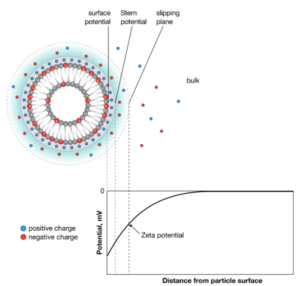
The zeta potential of a particle is the overall charge that a particle acquires in a particular medium, it has been defined as the potential at the hydrodynamic shear boundary. Larger zeta potentials predict a more stable dispersion, which means that all the particles in suspension will tend to repel each other thus preventing aggregation. Normally, particle suspensions with zeta potentials > +30 mV or < −30 mV are considered stable. Measurement of liposomes zeta potentials can provide insight about their stability, circulation times, protein interactions, particle cell permeability, and biocompatibility. In drug delivery system, specific zeta potential has the possibility to improve biological performance by circumventing surface charge related toxicities.
 Figure 1. Schematic representation of zeta potential. Immediately surrounding the liposome is a layer of tightly associated ions, opposite in charge to the surface of the sample. This is termed the Stern layer. Surrounding the Stern layer is a second layer of loosely associated ions. The point at which this second layer of ions moves with the liposome as a single entity is termed the slipping plane. Zeta potential is defined as the potential at this slipping plane, separating the liposome and associated ions from the ions of the bulk dispersing medium under an applied electric field. (Anal Bioanal Chem, 2017)
Liposome zeta potential determination methods
Electrophoresis-based method
Electrophoresis is used for estimating zeta potentials by applying an electric filed across the dispersion. Since the velocity is proportional to the magnitude of the zeta potential, the zeta potential can be calculated by detecting the migrate rate of particles toward the electrode of opposite charge.
Electroacoustic-based method
In this method, colloid vibration current and electric sonic amplitude are two main electroacoustic effects used in zeta potential characterization. These effects could be exploited to measure dynamic electrophoretic mobility in some commercial instruments to determine zeta potential. Electroacoustic techniques allow to achieve measurement in intact samples without dilution. In addition, densities and size are unnecessary information in electroacoustic-based method.
Creative Biostructure possesses advanced zeta potential analyzer to offer liposome zeta potential determination service. Our experts will take full consideration of varied parameters in working solutions, such as iron strength and pH. Please contact us for more information.
Ordering Process
References
- Smith MC, Crist RM, Clogston JD. (2017) Zeta potential: a case study of cationic, anionic, and neutral liposomes. Anal Bioanal Chem. 409: 5779-5787.
- Xu RL. (2008) Progress in nanoparticles characterization: sizing and zeta potential measurement. Particuology. 6: 112-115.
- TantraR, SchulzeP, QuinceyP. (2010) Effect of nanoparticle concentration on zeta-potential measurement results and reproducibility. Particuology. 8: 279-85.
