Liposome Visualization
Creative Biostructure has the most advanced Mempro™ Liposome technology, which can offer the best service for liposome manufacture and analysis. Based on our state-of-the-art equipment, we provide comprehensive characteristic determination service for clients worldwide.
Why visualization is important to liposome?
Liposomes have become widely used as pharmaceutical delivery carriers in numerous clinical applications. The increase in their applications has facilitated the development of analytical and technological approaches to determine their characteristics. Visualization analysis is an essential and powerful way for morphology studies, which allows intuitively imaging biological complex system at the molecular level. Through visualization analysis, a variety of useful data will be collected, including size distribution, bilayer organization, vesicles shape and number of lamellae. Liposome visualization data provided by microscopic techniques becomes prerequisite parameters guide the utilization of liposome for drug encapsulation.
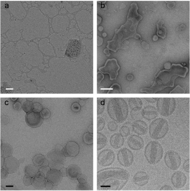
 Figure 1. Doxorubicine liposomes imaged by the most frequently used techniques in soft matter electron microscopy: dried sample without staining (a), UAc stained sample after two minutes of drying (b), negative stained sample (UAc) (c) and cryo-TEM (d). White scale bars represent 200 nm and black scale bars 50 nm. (Advanced science news, 2017)
Liposome visualization determination methods
Transmission electron microscopy (TEM)
TEM is a high-resolution imaging technique to evaluate morphology and architecture of liposomes. Several methods can be used to apply TEM including negative staining and flash-freeze. Negative staining TEM is a more convenient approach but with some staining artifacts that might interfere liposome internal structure. To avoid interference from NS-TEM, cryo-TEM is an optimal approach to study ultrastructure of rapidly frozen liposomes, but it requires extra caution and long time for sample preparation. Since sample preparation maintains the aqueous environment in cryo-TEM, it is particularly suitable for aqueous formulations such as protein solutions and liposomes.
Environmental scanning electron microscopy (ESEM)
ESEM has potential ability to image a liposome in its natural hydrated state, unlike other electron microscopic methods that often generate artifacts from staining, drying, fixation. It is the ideal scenario to visualize the formation and morphology of liposomes in real time. It also allows variation of the sample environment through a range of pressures, temperatures, and gas compositions. However, the resolution of ESEM is not high enough to study the surface properties and architecture of liposome.
Atomic force microscopy (AFM)
AFM achieves sample analysis in the wet mode, meanwhile, it allows the observation of the liposomal morphology without any sample manipulation such as staining labeling, or fixation. AFM also offers the unique opportunity to study the surface properties of liposome. However, liposomes might change their shape after deposition on mica due to the interaction between liposomes and mica, as well as the continuous tipping movement.
Confocal laser scanning microscopy (CLSM)
Labeling lipid bilayers using a fluorochrome marker, CLSM allows to acquire three-dimensional and high resolution images under variation environment to study liposome structure. Therefore, CLSM is a good method to study the deformability of liposome. Nevertheless, CLSM is limited by its native resolution which can only resolve structures under 200nm in diameter.
Creative Biostructure offers reliable protocols and a comprehensive set of equipment for liposome visualization at our analytical center. Please contact us to share more details about your research. Our tech specialists will provide professional consultation services and solutions to some of the toughest challenges.
Ordering Process
References
- Yvonne perrie, Afzal u. r. mohammed. (2007) Environmental scanning electron microscopy offers real-time morphological analysis of liposomes and niosomes. Journal of Liposome Research, 17:27-37.
- Barbara Ruozi, Daniela Belletti, Andrea Tombesi et al. (2011) AFM, ESEM, TEM, and CLSM in liposomal characterization: a comparative study. International Journal of Nanomedicine 6: 557-563.
- Linda E. Franken, Egbert J. Boekema, Marc C. A. Stuart. (2017). Transmission electron microscopy as a tool for the characterization of soft materials: application and interpretation. Adv. Sci. 4, 1600476.
