Nano-Flow Cytometry (nFCM) Service
Conventional flow cytometry is a rapid multi-parameter quantitative analysis technique for the physical and chemical properties of single cells or particles with cell size. By focusing the cells or particles in the suspension with hydrodynamics and passing through the laser detection area one by one, then the forward angle and multi-fluorescence signals can be detected by the detection system at the same time, so that the size of cells or particles can be qualitatively analyzed. However, it is difficult to characterize exosomes due to the small particle size, high heterogeneity, low refractive index, and low biomarker content. Nano-flow cytometry (nFCM) technology is an emerging method for nanoparticle characterization, which can detect exosomes with sizes as low as 30 nm. It provides a technical foundation for promoting exosome research in the field of clinical disease diagnosis and treatment.
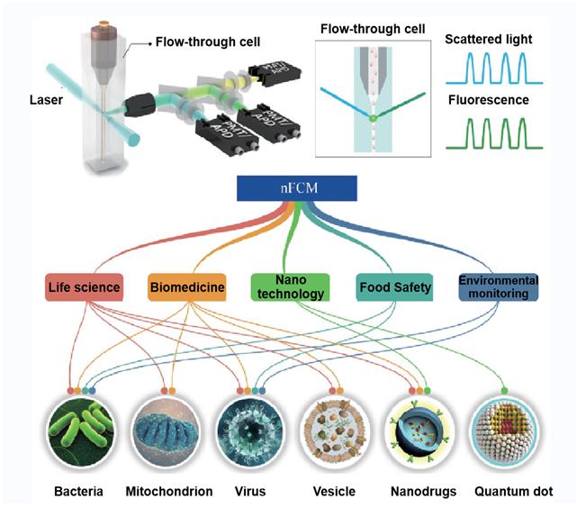 Figure 1. The application fields of nano-flow cytometry
Advantages of nanoparticle flow cytometry include the following aspects:
- High detection sensitivity: it can detect the nanoparticles with low refractive index and the fluorescence of single phycoerythrin.
- Multi parameters: it can simultaneously detect multi-parameters of a single nanoparticle and reveal a variety of biochemical properties of particles with multi-color fluorescence.
- Low sample size: it is suitable for the detection of precious samples.
- Fast speed: it can be comparable to TEM and other technologies.
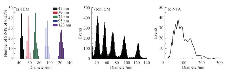 Figure 2. Comparison of characterization of SiNPs size distribution measured by TEM, nFCM, and NTA, respectively (Tian Y, et al. 2018)
At present, the nFCM technology can be applied to the following aspects:
- Detection of bacteria at single cell level
Through high-throughput and multi-parameter quantitative analysis at the single particle level, nFCM has been widely used in microbiology research, such as detecting pathogens in food, environmental water samples, and clinical samples and characterizing the stress response of bacteria to antibiotics, which has played a great role in promoting the research of food safety.
- Multi-parameter detection of mitochondria at the single organelle level
Compared with whole cell analysis or Western blot, nFCM can not only reveal the individual differences, but also simplify the complexity of samples and make it clear and concise for experimental data analysis.
- Detection of the virus at the single-particle level
By improving the laser power density of nFCM and further reducing the volume of detection area, the single particle scattering detection and small virus (MS2) with a particle size of only 27 nm can be realized. In addition, single particle detection makes nFCM insensitive to the influence of impurity particles, which is more suitable for the analysis of polydispersed or mixed samples and has been successfully applied to the purity determination of virus samples and the dynamic monitoring of the release process of the virus genome.
- Detection of EVs at the single-particle level
The particle size of most EVs is less than 100 nm, and the expression of specific proteins is very low and heterogeneous, which brings many challenges to the multi-parameter quantitation of EVs at the single particle level. nFCM shows significant advantages in the characterization of physical parameters like particle size distribution and concentration of EVs and biochemical properties such as nucleic acid. Because of its high lateral scattering detection sensitivity, nFCM can realize the single particle level multi-parameter quantitative detection of EVs with a particle size as low as 40 nm.
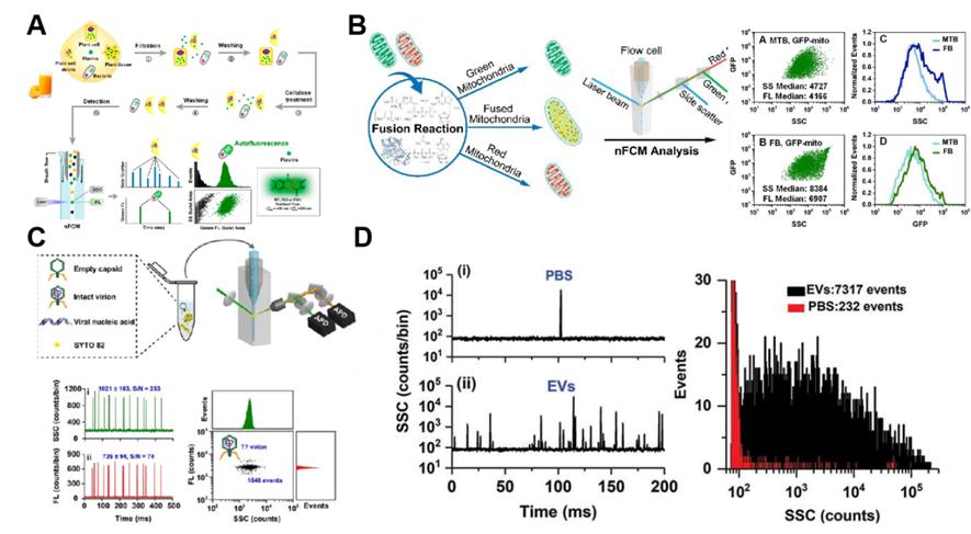 Figure 3. Detection of bacteria, mitochondria, virus and EV with nFCM (He S, et al. 2019)
In addition, nFCM can be used for the characterization of nanodrugs and single particle level quantum dots. With our exosome platform, Creative Biostructure can provide the nFCM service for different applications. If you are interested in our services, please feel free to contact us. We are looking forward to cooperating with you!
Ordering Process

References
- Tian Y, et al. Protein profiling and sizing of extracellular vesicles from colorectal cancer patients via flow cytometry. ACS nano. 2018. 12(1): 671-680.
- He S, et al. Label-free detection of bacteria in fruit juice by nano-flow cytometry. Analytical Chemistry. 2019. 92(3): 2393-2400.
- Luo X, et al. Activatable Mitochondria‐Targeting Organoarsenic Prodrugs for Bioenergetic Cancer Therapy. Angewandte Chemie International Edition. 2021. 60(3): 1403-1410.
- Niu Q, et al. Quantitative assessment of the physical virus titer and purity by ultrasensitive flow virometry. Angewandte Chemie. 2021. 133(17): 9437-9442.
- Tian Y, et al. Quality and efficiency assessment of six extracellular vesicle isolation methods by nano-flow cytometry. Journal of Extracellular Vesicles. 2020. 9(1): 1697028.

