Exosomes Isolated from Animal Plasma
The content of exosomes reflects the originating cell type and its physiological or pathological state, providing unique insights into cellular activities and systemic processes. Among the various biological fluids from which exosomes can be isolated, animal plasma, which serves as the fluid component of blood, is a particularly rich source due to its high abundance of exosomes derived from various cell types. The increasing interest in plasma-derived exosomes for biomedical research is driven by their unique properties, such as stability in the circulation, ability to cross biological barriers, and potential to carry diverse cargoes that reflect the state of the organism.
Creative Biostructure offers various exosomes isolated from animal plasma, including exosomes isolated from bovine plasma, canine beagle plasma, and more as standard research controls to support your veterinary disease research.
Product List
Exosomes Isolated from Animal Plasma: Accessibility and Systemic Insight
Exosomes are secreted by nearly all cell types and are found in various biological fluids including plasma, urine (HQExo™ Exosome-urine), saliva (HQExo™ Exosome-saliva) and cerebrospinal fluid (HQExo™ Exosome-CSF). Exosomes carry multiple membrane-bound, biologically active molecules, and their molecular cargo mimics the surface molecular profile of their cell of origin. The vesicular contents of exosomes include nucleic acids, enzymes, soluble factors, and a variety of molecules derived from the cytosol of the cell of origin. Their ability to transfer this cargo to other cells enables them to play an essential role in intercellular signaling, disease progression and therapeutic responses.
In veterinary preclinical and clinical studies, exosomes from animal plasma have the potential to significantly improve diagnostic capabilities and therapeutic approaches. These exosomes can be used to identify novel biomarkers for early detection of disease, enabling more accurate and timely diagnosis. In addition, exosomes hold promise for the development of targeted therapies as they can carry proteins, lipids and RNA that influence disease pathways.
Exosomes isolated from plasma are particularly useful for research because of several unique characteristics. First, animal plasma is a readily accessible and minimally invasive biological fluid that allows for repeated sampling without harming the organism. This accessibility is particularly advantageous in veterinary preclinical research, where the study of disease progression and the evaluation of novel therapeutics often require frequent sampling.
Second, plasma-derived exosomes reflect the systemic state of the organism, providing a snapshot of cellular activity in different tissues and organs. The diverse cell types that secrete exosomes into the plasma, including immune cells, endothelial cells, and tumor cells, contribute to the complexity and breadth of the exosomal cargo. This feature makes plasma-derived exosomes an ideal candidate for the study of intercellular communication, as they can reflect the interactions between different biological systems and provide insights into animal disease states.
Isolation and Characterization of Exosomes from Animal Plasma
The isolation of exosomes from plasma presents several challenges. Plasma contains exosomes from different cell types in varying proportions, making it difficult to identify their specific origin. Exosomes are often coated with proteins, glycoproteins, or glycolipids, which can cause aggregation and potential loss during centrifugation. The presence of other vesicles, protein complexes, and subcellular fragments in plasma complicates the isolation of pure exosomal fractions. The propensity of exosomes to form aggregates of various sizes can lead to losses and further complicate their recovery. These factors make the isolation of well-defined exosomal fractions from human plasma a challenging task.
Nevertheless, several methods have been developed to isolate exosomes from plasma, including ultracentrifugation, size exclusion chromatography, and precipitation-based methods.
Ultracentrifugation
Ultracentrifugation is the gold standard for isolating exosomes from plasma. The process involves high-speed centrifugation that separates exosomes from other plasma components based on their size and density (e.g., density gradient ultracentrifugation). Typically, plasma is first subjected to low-speed centrifugation to remove larger particles such as cells and debris. The supernatant is then subjected to higher-speed centrifugation to pellet the exosomes, which can be further purified by additional gradients or filtration steps. Ultracentrifugation provides high yield and purity, but is time consuming and requires specialized equipment.
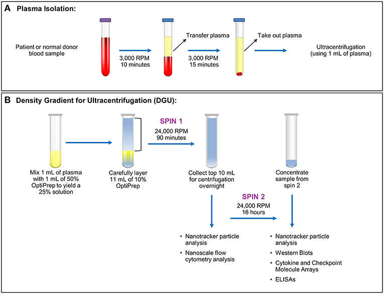 Figure 1. Plasma exosome isolation by density gradient ultracentrifugation (DGU). (Cumba Garcia et al., 2019)
Size-Exclusion Chromatography (SEC)
Size-exclusion chromatography separates exosomes based on their size as they pass through a porous medium. Larger particles, such as exosomes, elute faster than smaller molecules, allowing for efficient isolation. SEC is considered less harsh than ultracentrifugation and may preserve the biological activity of exosomes. However, the yield can be lower than with ultracentrifugation and the technique can be more time-consuming.
Precipitation Reagents
Precipitation-based methods (polymer precipitation) use proprietary reagents to induce aggregation of exosomes, which are then collected by centrifugation. These methods are inexpensive, easy to implement, and can process large volumes of plasma. However, the purity of the exosomes may be lower than other methods because this technique often co-isolates proteins and other contaminants.
Characterization Methods
Once isolated, exosomes are typically characterized using a combination of techniques, including:
- Nanoparticle Tracking Analysis (NTA): Used to determine the size distribution and concentration of exosomes.
- Transmission Electron Microscopy (TEM): Provides high-resolution images of exosome morphology.
- Flow Cytometry: Allows for the analysis of exosome surface markers and characterization of exosome subpopulations.
- Western Blotting: Used to identify exosome-specific markers such as CD63, CD9, and TSG101.
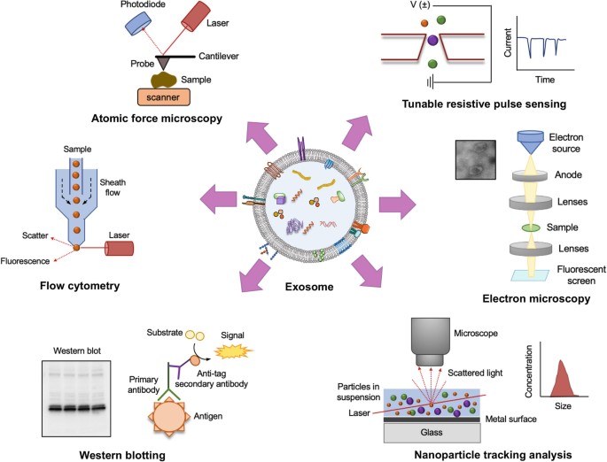 Figure 2. Methods for characterization of exosomes in biological samples (Singh et al., 2024)
Applications of Exosomes Isolated from Animal Plasma
Veterinary Preclinical and Clinical Studies
Exosomes isolated from animal plasma play a significant role in veterinary preclinical and clinical studies. They are used for disease modeling, enabling the study of animal-specific diseases and their progression. They also serve as non-invasive biomarkers for monitoring health conditions, disease states, and therapeutic responses in animals. In addition, animal plasma exosomes are valuable for testing novel drugs and therapies because they provide a more accurate representation of systemic biological processes than cell line-derived exosomes. The use of standard plasma exosomes with disease samples facilitates the investigation of veterinary preclinical and clinical studies.
Intercellular Communication Studies
Plasma exosomes mediate communication between different cell types in different tissues. Studying these exosomes can provide valuable insights into how cells in the body interact with each other, which is important for understanding immune responses, tissue regeneration, and inflammation.
Recent Case Studies on Exosomes Isolated from Animal Plasma
Case Study 1: Systematic analysis of different degrees of hemolysis on miRNA levels in serum and serum-derived extracellular vesicles from dogs
This study investigates the effect of hemolysis on circulating microRNAs (miRNAs) in dogs, focusing on serum and serum-derived extracellular vesicles (EVs). In a controlled in vitro experiment, hemolysis significantly altered the levels of miR-16, miR-92a, miR-191, miR-451, and miR-486, while other miRNAs remained unaffected. The study also found that some canine miRNA abundances differ from those reported in humans. These results are the first to describe the effect of hemolysis on circulating miRNAs in dogs and highlight the importance of accounting for hemolysis in plasma- or serum-based miRNA studies.
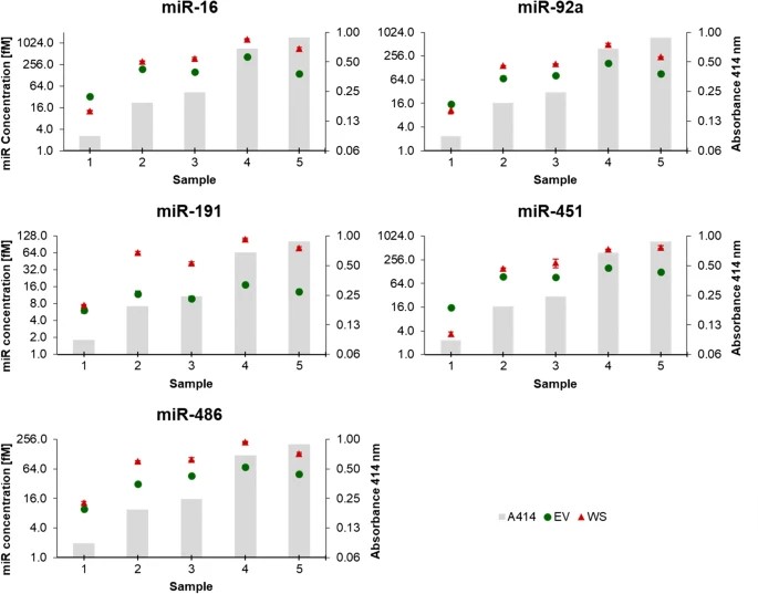 Figure 3. Absolute quantification of miRNAs sensitive to haemolysis in whole serum (WS) and serum-derived extracellular vesicles (EV) groups, in patients' sera with different degrees of haemolysis (validation experiments). Grey columns in the background depict increasing levels of haemolysis, measured using the haemoglobin absorbance at 414 nm. (Aguilera-Rojas et al., 2022)
Case Study 2: Plasma exosomes regulate systemic blood pressure in rats
This study investigates the role of plasma exosomes in the regulation of systemic blood pressure and cardiovascular function in normotensive and hypertensive rats. Exosomes, small extracellular vesicles containing various molecules, are known to mediate cell communication and influence physiological and pathological processes, including hypertension. The researchers hypothesized that plasma-derived exosomes could affect blood pressure regulation in normotensive Wistar Kyoto rats (WKY) and spontaneously hypertensive rats (SHR).
Exosomes were isolated from plasma and administered intraperitoneally to both groups of rats for six weeks. The results showed that SHR-derived exosomes increased systolic blood pressure in WKY rats, whereas WKY-derived exosomes decreased blood pressure in SHR. In addition, SHR-derived exosomes induced structural changes in the thoracic aorta of WKY rats, such as wall thickening and decreased collagen abundance, similar to the changes observed in SHR. Conversely, WKY-derived exosomes helped to reverse these changes in SHR.
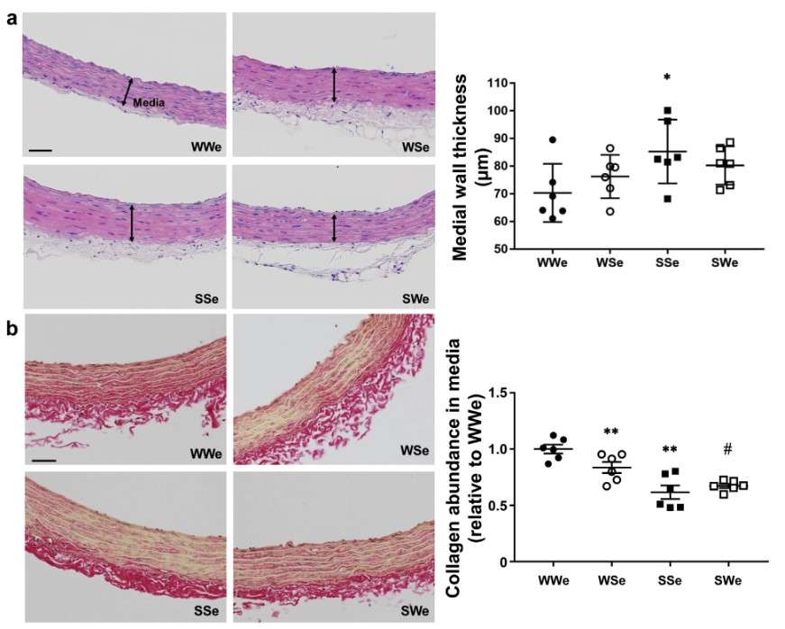 Figure 4. Effects of plasma exosomes on wall thickness and medial collagen abundance of thoracic aorta in WKY and SHR. (a) Representative hematoxylin and eosin-stained sections (4 mm) for thoracic aorta isolated from WKY and SHR (10-week-old) treated with plasma exosomes weekly for 6 weeks were shown (n = 6). Medial wall thickness of aorta was calculated and shown as means ± SEM (dot plot graph). (b) Representative picrosirius red stained sections (4 mm) for thoracic aorta from WKY and SHR (10-week-old) treated with plasma exosomes weekly for 6 weeks were shown (n = 6). Collagen was stained in red. The intensity of red region was calculated as collagen abundance, normalized to perimeter of aorta and shown as means ± SEM (dot plot graph). Scale bar: 50 mm * P < 0.05 vs. WWe; ** P < 0.01 vs. WWe; # P < 0.05 vs. SSe. (Otani et al., 2018)
Advantages of Our Exosomes Isolated from Animal Plasma
- Abundance and Accessibility: Exosomes isolated from plasma are abundant and easily accessible, making them an ideal source for research. This accessibility allows for large-scale collection and analysis, which is critical for both veterinary preclinical and clinical research.
- Diverse Cell Source Representation: Plasma-derived exosomes can be derived from a wide variety of cell types, providing a broad spectrum of molecular information. The ability to isolate exosomes from different animal models allows researchers to study the effects of exosome-based therapies in species-specific contexts, facilitating the development of cross-species applications.
- Broad Applicability: Exosomes from various animal species can be used to study a wide range of animal diseases, including cancer, cardiovascular disease, autoimmune disease and infectious diseases.
Exosome Resources


FAQs About Exosomes Isolated from Animal Plasma
-
What are exosomes, and how are they isolated from plasma?
Exosomes are small vesicles secreted by cells that contain various biomolecules, including proteins, lipids, and RNA. They are isolated from plasma through a series of steps, including blood collection, centrifugation, and purification techniques such as ultracentrifugation or size-exclusion chromatography.
-
What are some applications of plasma-derived exosomes?
Plasma-derived exosomes are valuable for disease modeling, biomarker discovery, therapeutic delivery, and intercellular communication studies. Enhance your research by comparing our high-quality standards to your samples for more accurate insights.
-
Which downstream analyses can your products be used?
HQExo™ standard exosomes could use as positive controls for exosome isolation and functional research, such as nanoparticles tracking analysis (NTA), flow cytometry, ELISA, and Western blotting.
-
Do you offer exosomes derived from human plasma?
Yes, in addition to exosomes isolated from animal plasma, we also provide exosome derived from human plasma (healthy donors) (HQExo™ Exosome-Plasma). These are available on the Exosomes Isolated from Body Fluids.
Creative Biostructure's standard exosome products ensure higher purity and quality to meet our customers' research needs. Contact us today to learn more about how our animal plasma exosome products can enhance veterinary disease studies.
References
- Aguilera-Rojas M, Sharbati S, Stein T, Candela Andrade M, Kohn B, Einspanier R. Systematic analysis of different degrees of haemolysis on miRNA levels in serum and serum-derived extracellular vesicles from dogs. BMC Vet Res. 2022;18(1):355.
- Cumba Garcia LM, Peterson TE, Cepeda MA, Johnson AJ, Parney IF. Isolation and analysis of plasma-derived exosomes in patients with glioma. Front Oncol. 2019;9:651.
- Garcia-Contreras M, Shah SH, Tamayo A, et al. Plasma-derived exosome characterization reveals a distinct microRNA signature in long duration Type 1 diabetes. Sci Rep. 2017;7(1):5998.
- Muller L, Hong CS, Stolz DB, Watkins SC, Whiteside TL. Isolation of biologically-active exosomes from human plasma. Journal of Immunological Methods. 2014;411:55-65.
- Otani K, Yokoya M, Kodama T, et al. Plasma exosomes regulate systemic blood pressure in rats. Biochemical and Biophysical Research Communications. 2018;503(2):776-783.
- Singh S, Dansby C, Agarwal D, Bhat PD, Dubey PK, Krishnamurthy P. Exosomes: methods for isolation and characterization in biological samples. In: Di Nardo P, Dhingra S, Desiderio V, eds. Adult Stem Cells: Methods and Protocols. Springer US; 2024:181-213.

