Exosome Isolation by Size-exclusion Chromatography (SEC)
Exosome isolation is a critical step in research, and size exclusion chromatography (SEC) has emerged as an effective and gentle technique for exosome preparation. This method is attractive for studying exosome biology, diagnosing diseases, and developing new treatment strategies.
As a leader in exosome research, Creative Biostructure offers SEC technology to isolate exosomes with high yield and purity, assisting clients in further exploring the significant potential of exosomes in diagnosis and treatment.
What is Size Exclusion Chromatography?
SEC separates molecules according to their size through gel filtration. In this method, the gel consists of spherical beads containing pores of a specific size. When molecules of different sizes enter or are eliminated into these pores, separation occurs. Small molecules can diffuse into the pores and are delayed in flowing within the column due to their size, while large molecules cannot enter the pores and will wash out in the volume of the column voids. Therefore, gradually elute and separate in molecular weight (MW) order when passing through the chromatographic column.
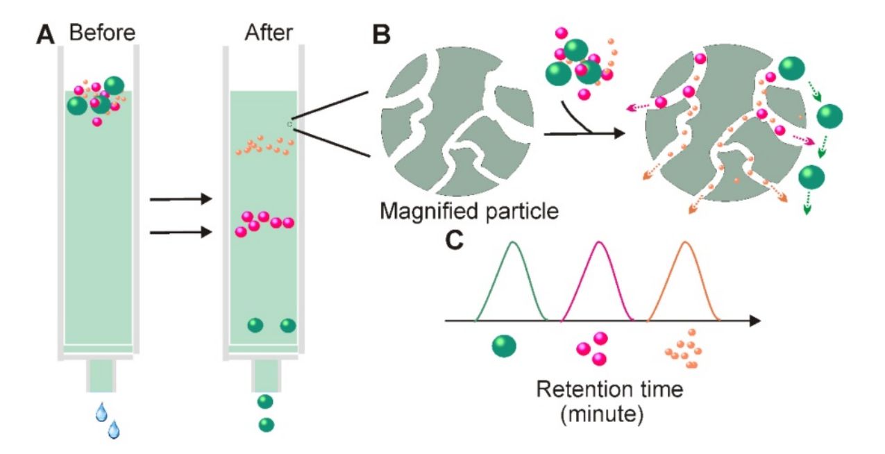 Figure 1. Schematic of size exclusion chromatography–based exosome isolation. (Yang D, et al., 2020)
How Does Size Exclusion Chromatography Work?
| Process | Details |
| a) Sample preparation | The sample is usually clarified by centrifugation or filtration to remove large fragments and cellular components. |
| b) Column selection | The selection of an appropriate SEC column is essential for efficient separations. The choice of column depends on the size range of the exosomes. |
| c) Column equilibrium | Before loading the sample, the SEC column must be equilibrated with an appropriate buffer or mobile phase to prevent unwanted interactions between the sample and the column matrix. |
| d) Sample loading | Introduce the sample into the column manually or using an automated system. The amount of sample added should be within the capacity of the column. |
| e) Elution | After sampling, particles in the sample migrate through the porous beads depending on their size as the mobile phase flows through the column. |
| f) Fraction collection | Monitor the elution curve by detecting and recording eluted particles. Collect scores at specific time intervals or based on detecting particles of interest. |
| g) Characterization and analysis | Further analyze the collected fractions to confirm the presence of exosomes or target particles. |
What are the General Precautions for Size Exclusion Chromatography?
- Particles and pore size – Generally speaking, the smaller the particle size, the higher the resolution. The exclusion limit and classification range of aperture-controlled media.
- Chromatographic column size - The resolution increases with the length of the chromatographic column and the diameter of the chromatographic column.
- Column packing - Column packing is crucial for separation, and overfilled chromatography columns may cause the pores in the beads to collapse, leading to a decrease in resolution. Underfilled chromatographic columns increase the mixing volume outside the pores, resulting in wider peaks and lower resolution.
- Flow rate - Medium flow rate provides the highest resolution. A medium flow rate allows molecules time to fully enter the surface area of the stationary phase, allowing smaller molecular weight substances time to enter the pores, thereby improving the distribution of substances with different molecular weights.
Besides Isolation, What are the Other Applications of SEC in Exosome Research?
- Characterization: SEC can be used to analyze the size distribution of exosomes in a sample, which is essential for understanding the biological function of exosomes and downstream functional research.
- Biomarker discovery: SEC is used to isolate and enrich specific subpopulations of exosomes, enabling the identification of potential biomarkers for various diseases.
- Drug delivery: By loading therapeutic molecules into exosomes and purifying them using SEC, researchers can create highly targeted and efficient drug delivery systems for a variety of diseases, including cancer and inflammation.
In addition to SEC-based exosome isolation services, we help clients explore the role of exosomes in intercellular communication, immune response regulation, and disease progression through a range of high-purity exosome products.
| Cat No. | Product Name | Source |
| Exo-HDBF-01 | HQExo™ Exosome-SDH-Alzheimer's plasma | Exosome derived from Single Donor Human Alzheimer's plasma |
| Exo-HDBF-02 | HQExo™ Exosome-SDH-Asthma plasma | Exosome derived from Single Donor Human Asthma plasma |
| Exo-HDBF-03 | HQExo™ Exosome-SDH-Atopic Dermatitis plasma | Exosome derived from Single Donor Human Atopic Dermatitis plasma |
| Exo-HDBF-04 | HQExo™ Exosome-SDH-Benign Breast Conditions plasma | Exosome derived from Single Donor Human Benign Breast Conditions plasma |
| Exo-HDBF-05 | HQExo™ Exosome-SDH-Benign Organ Tumor plasma | Exosome derived from Single Donor Human Benign Organ Tumor plasma |
| Exo-HDBF-06 | HQExo™ Exosome-SDH-Benign Prostatic Hyperplasia plasma | Exosome derived from Single Donor Human Benign Prostatic Hyperplasia plasma |
| Explore All Exosomes Isolated from Human Disease-state Body Fluids | ||
Why Choose Size Exclusion Chromatography?
- Mild and non-destructive
- Multifunctionality and flexibility
- Relatively fast and efficient
- Widely applicable and scalable
SEC is the best method for isolating exosomes from most proteins while recovering intact and functionally intact exosomes from plasma. According to reports, SEC technology is particularly useful in exosomes derived from adipose tissue.
Creative Biostructure is delighted to be your professional partner in the field of exosomes and is willing to provide you with first-class exosome isolation services and high-quality exosome products. Please feel free to contact us for a detailed quote.
References
- Yang D, et al. Progress, opportunity, and perspective on exosome isolation - efforts for efficient exosome-based theranostics. Theranostics. 2020. 10(8): 3684-3707.
- Liu WZ, et al. Current status and outlook of advances in exosome isolation. Anal Bioanal Chem. 2022. 414(24): 7123-7141.
- Sidhom K, et al. A Review of Exosomal Isolation Methods: Is Size Exclusion Chromatography the Best Option?. Int J Mol Sci. 2020. 21(18): 6466.

