Mempro™ Virus-like Particles (VLPs) in Bioimaging
Creative Biostructure has developed an unparalleled and engineering virus-like particles (VLPs) technology platform in recent years. We have made a lot of efforts on VLPs production and application to facilitate the membrane protein research and biomedicine development. Bioimaging is one of our major application of VLPs. Two advantages of VLPs for bioimaging are their ability to genetic or chemical conjugation and their great biological compatibility.
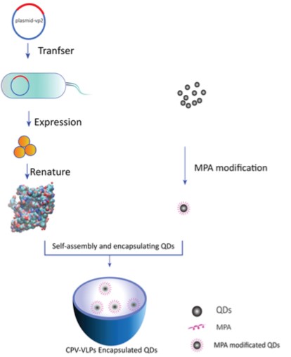
 Figure 1. Schematic of encapsulation of QDs with CPV-VLPs for bioimaging. (Plos One, 2015)
Mempro™ Virus-like Particles (VLPs) platform has already established a broad range of chemical engineering and genetic methods to allow VLPs to load large amount of imaging reagents such as fluorescent dyes or probe. To give an example, fluorescent CPMV sensors are able to make visualization of vasculature and blood flow in living cells possible, and these sensors can be used to image tumor angiogenesis in a variety of human cancer cell lines.
Another strategy for VLPs bioimaging applications is to design hybrid VLPs with fluorescent cores (such as quantum dots and cyanine dye) by our advanced encapsulation technology, which allowing confocal microscopy tracking and infrared optical imaging. Moreover, this strategy only used inside part of VLPs to encapsulate the imaging reagents, making the outside part of VLPs still available for chemical modifications.
Furthermore, non-invasive imaging methods such as MRI and PET have applied VLPs as imaging reagents as well. For example, Creative Biostructure can modified our custom VLPs with gadolinium to be MRI contrast reagents because VLPs have large surface areas to be decorated, and the rigid structures and large rotational correlation of VLPs provide high relaxivity values.
GFP and Quantum dots (QDs) were widely applied for in vivo and in vitro imaging as alternatives to labeling. For example, fluorescent cVLPs (chimeric virus-like particles) of canine parvovirus can be expressed in insect cells. In order to create the fluorescent cVLPs of canine parvovirus, GFP was genetically engineered onto the N-terminal region of the viral protein VP2, as a visualization tool to understand mechanisms of viral infections. GFP was also used to design chimeric HIV VLPs allowing protein to be followed during assembly and transmission using live-cell imaging.
Our VLPs platform also provide a variety of biomedical applications such as diagnosis, monitoring, therapy and disease prevention. Mempro™ Virus-like Particles (VLPs) Application services in vaccines, cell targeting, delivery systems, and lipoparticle technology are also available. Please feel free to contact us for a detailed quote.
References:
D. Yan, et al. (2015). Quantum Dots Encapsulated with Canine Parvovirus-Like Particles Improving the Cellular Targeted Labeling. Plos One,10(9): e0138883.
I. Yildiz, et al. (2011). Applications of viral nanoparticles in medicine. Curr. Opin. Biotechnol., 22(6): 901-908.
K. Li, et al. (2010). Viruses and their potential in bioimaging and biosensing applications. Analyst, 135: 21-27.