Labeling Exosomes with Gold Nanoparticles (GNPs)
Exosomes are cell-derived lipid bilayer-enclosed nanovesicles that exchange various components with neighboring and distant cells through the extracellular environment, thereby participating in physiological and pathological processes such as wound healing, immune responses, and tumorigenesis. These properties of exosomes provide opportunities for diagnostic and therapeutic innovations. Consequently, there is a growing interest in the clinical application of exosomes as biomarkers and for therapeutic purposes.
To successfully translate exosomes to the clinic, it is essential to understand their in vivo behavior. Advances in molecular imaging have enabled high-resolution tracking of exosomes, providing valuable information on exosome transport, biodistribution, cellular uptake, and molecular mechanisms. A prerequisite for performing tracking experiments is exosome labeling. Gold nanoparticles (GNPs) are emerging as a promising material that can be easily applied to label exosomes.
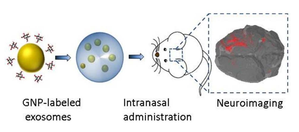 Figure 1. In vivo neuroimaging of exosomes using gold nanoparticles. (Betzer O, et al., 2017)
What are GNPs?
Computerized tomography (CT), a non-invasive imaging method that takes images of deep structures in the body, may be useful for exosome tracking in vivo. However, exosomes cannot be detected directly by CT, and a contrast agent must be added to label the exosomes before they can be visualized. Advances in nanotechnology have enabled the use of ultra-small GNPs for exosome labeling. The GNPs have received much attention as CT contrast agents because they are easy to synthesize and surface-functionalize, have strong X-ray attenuation, a high degree of flexibility in coating and targeting functional groups, and are non-toxic and biocompatible in vivo. The use of GNPs for cell labeling and tracking of labeled cells using CT has become a time-saving and cost-effective method. Active targeting of GNPs can enhance contrast and sensitivity and further reduce the radiation dose required during CT imaging.
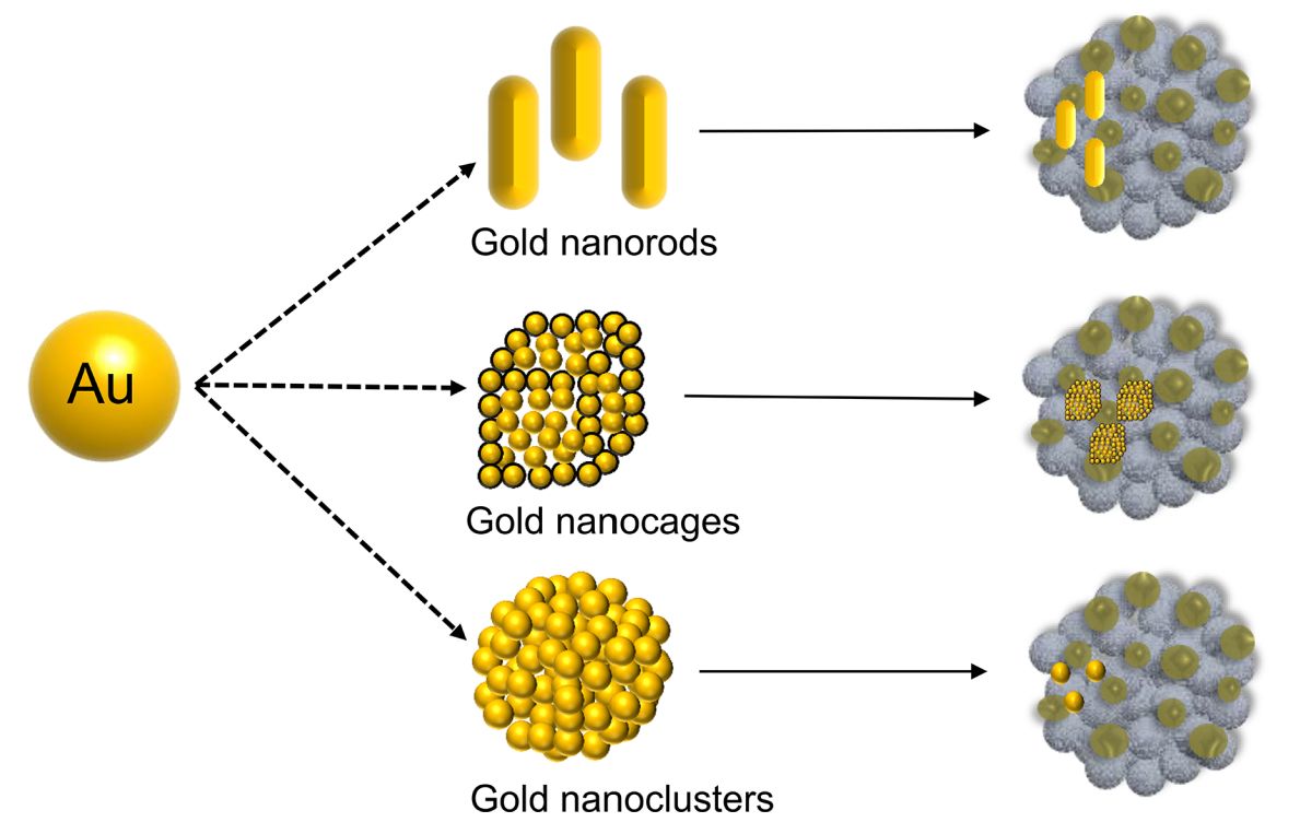 Figure 2. Different shapes of GNPs. (Yang Z, et al., 2022)
How do GNPs Conjugate with Exosomes?
1. Freshly prepared citrate-terminated gold nanoparticles (CGNP) are polyethylene glycolized (incubated with PEG) to form polyethylene glycolized gold nanoparticles (PGNP).
2. The PGNP are functionalized with antibodies corresponding to exosome surface proteins and finally incubated with human serum, in which the affixation between CGNP and exosomes occurred.
3. After centrifugation, the PGNP-exosome complex settles to the bottom of the tube.
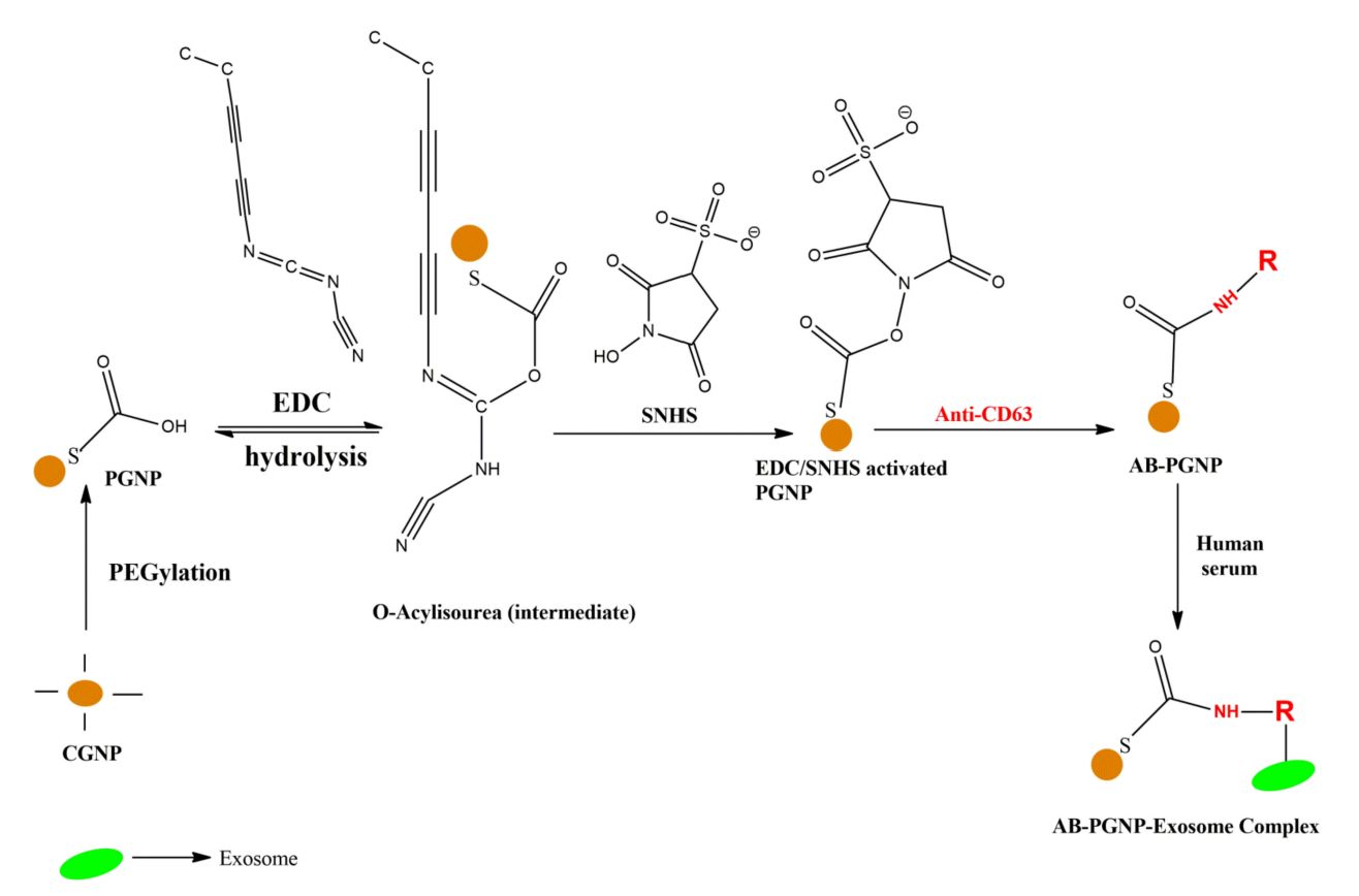 Figure 3. A brief workflow of the conjugation of gold nanoparticles with exosomes. (Pammi Guru KT, et al., 2022)
Advances in Research on Labeling Exosomes with GNPs
An indirect strategy is usually used to label exosomes. GNPs are added to the parental cell culture medium and taken up by the cells through endocytosis. Some contrast agents may be loaded onto exosomes during their biogenesis. However, this indirect strategy results in only a small fraction of GNPs being loaded onto exosomes. Some direct strategies introduce GNPs by transiently disrupting the exosome membrane, but this may compromise the integrity and functionality of the exosome.
In recent years, researchers have established a non-invasive in vivo method for neuroimaging and tracking exosomes based on labeling of glucose-encapsulated GNPs and computed tomography imaging. The research demonstrated that glucose-encapsulated GNPs mediate endocytosis into MSC exosomes via the glucose transporter protein GLUT-1. This research further determined the optimal GNP size as well as the mode of administration. This exosome labeling technique could serve as a powerful diagnostic tool for a variety of brain disorders and may potentially enhance exosome-based neuronal restoration therapies.
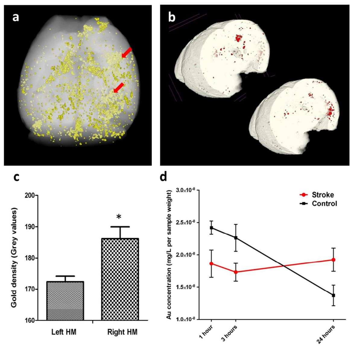 Figure 4. Ex vivo imaging and gold quantification within the brain. (Betzer O, et al., 2017)
What are the Advantages of Labeling Exosomes with GNPs?
- Target Specificity - When exosomes are labeled with GNPs, they can be specifically targeted to certain tissues or cells. This is particularly useful for drug delivery systems, which aim to deliver drugs specifically to diseased cells while minimizing damage to surrounding healthy cells.
- Sensitive and Accurate Detection - GNPs have unique optical properties that allow them to be used in a variety of sophisticated detection methods, including colorimetric assays, spectroscopic measurements, and more. Since exosomes are tiny vesicles, combining them with GNPs can increase sensitivity and thus the accuracy of the assay.
- Enhanced Therapeutic Effects - In some cases, GNPs themselves can enhance the therapeutic effects of exosomes. For example, GNPs can enhance the heating effect of cancer cells upon irradiation, thereby effectively treating cancer.
Related Products
We are committed to providing one-stop exosome research services, including exosome isolation, characterization, labeling, tracking, in vivo, and in vitro functional analysis to explore their potential applications as cancer diagnostic markers and drug carriers. In addition, we provide high-quality exosome kits and exosome products for clients to choose from.
| Cat No. | Product Name | Source |
| Exo-CH04 | HQExo™ Exosome-H196 | Exosome derived from Human small cell lung cancer cell line (H196 cell line) |
| Exo-CH05 | HQExo™ Exosome-H841 | Exosome derived from human small cell lung cancer cell line (H841 cell line) |
| Exo-IC06 | HQExo™ Exosome-NK | Exosome derived from human NK cell line |
| Exo-IC04 | HQExo™ Exosome-RAWS 264.7 | Exosome derived from mouse macrophage cell line (RAWS 264.7) |
| Exo-HDBF-03 | HQExo™ Exosome-SDH-Atopic Dermatitis plasma | Exosome derived from Single Donor Human Atopic Dermatitis plasma |
| Exo-HDBF-04 | HQExo™ Exosome-SDH-Benign Breast Conditions plasma | Exosome derived from Single Donor Human Benign Breast Conditions plasma |
| Exo-SC03 | HQExo™ Exosome-hTERT | Exosome derived from hTERT-immortalized Mesenchymal Stem Cell |
| Exo-SC04 | HQExo™ Exosome-MSC | Exosome derived from Xeno-Free Human Mesenchymal Stem/Stromal Cells and Media |
| Explore All Exosome Products | ||
Creative Biostructure's GNPs-based labeling is an effective tool for researching exosome characterization and transport for therapeutic and diagnostic applications. If you are interested in exosome labeling services, please do not hesitate to contact us for more information.
References
- Betzer O, et al. In Vivo Neuroimaging of Exosomes Using Gold Nanoparticles. ACS Nano. 2017. 11(11): 10883-10893.
- Yang Z, et al. The Applications of Gold Nanoparticles in the Diagnosis and Treatment of Gastrointestinal Cancer. Front Oncol. 2022. 11: 819329.
- Pammi Guru KT, et al. Novel Gold Nanoparticle-Based Quick Small-Exosome Isolation Technique from Serum Sample at a Low Centrifugal Force. Nanomaterials. 2022. 12(10): 1660.
- Gong L, et al. In vivo CT imaging of gold nanoparticle-labeled exosomes in a myocardial infarction mouse model. Ann Transl Med. 2021. 9(6): 504.
- Chandrasekaran R, et al. Labeling and tracking cells with gold nanoparticles. Drug Discov Today. 2021. 26(1): 94-105.

