Single-crystal X-ray diffraction (XRD) is an advanced analytical technique that provides detailed insights into the atomic and molecular structure of crystalline materials. Through the diffraction of X-rays by a single crystal, XRD allows scientists to precisely determine the three-dimensional arrangement of atoms within a compound, including lattice parameters, symmetry, atomic positions, bond lengths, bond angles, configurations, and even electronic density distributions. This technique is crucial for understanding the structure of a wide range of materials, from small organic molecules to large biomolecules and complex inorganic compounds.
Definition and Basic Principles of XRD
Single-crystal X-ray diffraction works on the principle that when X-rays are directed at a crystal, they interact with the regularly spaced atoms in the crystal lattice. This interaction causes the X-rays to diffract in specific directions based on the atomic arrangement, creating a diffraction pattern. By analyzing these patterns, scientists can infer critical structural information about the crystal, such as bond lengths, bond angles, and the overall molecular geometry.
The process of XRD involves collecting data as the crystal is rotated to capture diffraction from various orientations. From this information, the exact three-dimensional structure of the compound can be determined, which is invaluable for understanding its chemical and physical properties.
Key Components of Single-Crystal XRD Equipment
The setup of a single-crystal X-ray diffraction experiment includes several key components:
- Rotating Target: The source of X-rays, which are typically generated by a high-voltage generator and focused onto the crystal.
- Angle Measurement Instrument: This device measures the angles at which X-rays are diffracted by the crystal.
- Detector: The detector captures the diffracted X-rays, converting the data into a readable form for analysis.
- Control/Processing System: The system that manages the experiment and processes the diffraction data.
- Cooling System: Keeps the crystal at a stable temperature during analysis, preventing thermal degradation.
The X-ray beam passes through the crystal and diffracts based on the crystal's atomic arrangement. The diffraction pattern is then analyzed using computational tools to extract detailed structural data, including bond lengths, angles, and atomic coordinates.
How Single-Crystal XRD Works in Practice
Crystal Preparation and Mounting
For accurate results, it is crucial to have high-quality crystals for single-crystal X-ray diffraction experiments. Crystals should be large, well-formed, and free of defects. Ideal crystals range from 30 to 300 microns in size. After preparation, the crystal is carefully mounted on a thin glass fiber using epoxy or other materials that do not interfere with diffraction.
The crystal must be placed in such a way that it can be rotated freely during the experiment, allowing diffraction data to be collected from all possible angles. A stable environment is also essential to prevent any external factors, such as temperature fluctuations, from affecting the results.
X-ray Source Selection and Diffraction Data Collection
Two primary X-ray sources are commonly used in single-crystal XRD experiments: copper (Cu) and molybdenum (Mo) targets.
- Cu Light Source: Cu has a scattering intensity 6-10 times greater than Mo, making it ideal for collecting data from small or weakly scattering crystals. However, Cu has a higher absorption effect, which can impact larger or denser crystals.
- Mo Light Source: Mo is particularly beneficial for studying structures that contain heavy atoms, such as those in metal-organic frameworks or proteins. Mo light sources are often preferred in protein crystallography because they provide clearer, more reliable diffraction patterns. Please read our article A Beginner's Guide to Protein Crystallography to get more information.
Data Processing and Software for Analysis
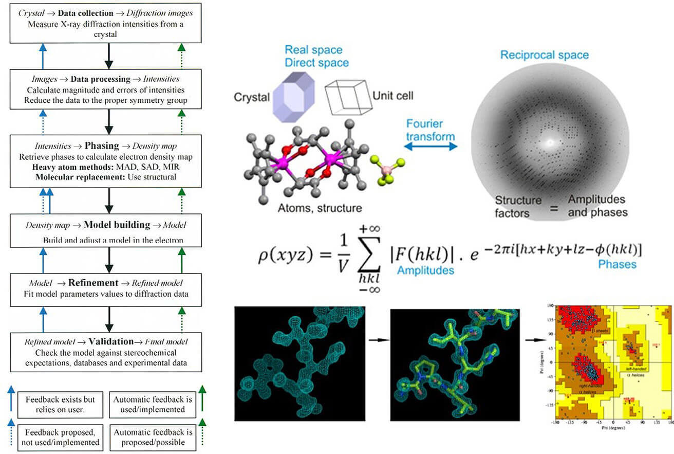
The data collected during the diffraction process is then analyzed using advanced computational tools and software. These programs perform tasks such as phase determination and refinement of the atomic model. Notable software tools include SHELXL and Olex2, which are widely used for structure refinement and visualization in X-ray crystallography.
Applications of Single Crystal X Ray Crystallography
Structural Determination of Organic Compounds
X-ray diffraction is frequently employed in chemical research to determine the structure of organic compounds, especially in the development of new drugs. By analyzing the diffraction patterns, scientists can deduce the three-dimensional structure of complex organic molecules, including the arrangement of atoms, bond lengths, and angles. This information is vital for understanding how these molecules interact in biological systems.
Analysis of Inorganic and Metal-Organic Frameworks
In the field of material science, single-crystal X-ray diffraction is essential for studying inorganic compounds and metal-organic frameworks (MOFs). These materials have applications in catalysis, energy storage, and gas adsorption. XRD allows scientists to precisely analyze the arrangement of metal ions and organic ligands within the framework, aiding the design of new materials with specific properties for targeted applications.
Advancements in Drug Design and Pharmaceutical Research
Single-crystal X-ray diffraction plays a critical role in pharmaceutical research by providing high-resolution structural data on drug-target interactions. By determining the precise binding sites and interactions between a drug molecule and its target protein, XRD helps guide the design of more effective and selective therapeutic agents. This structural information accelerates the process of drug discovery and optimization.
Case Studies in Single Crystal X-ray Crystallography
Case Study 1: Phase Identification of High-Entropy Perovskite Single Crystals via SC-XRD
A notable case study in the application of Single-Crystal X-ray Diffraction (SC-XRD) is the investigation of high-entropy perovskite single crystals. Researchers developed a method for synthesizing novel high-entropy semiconductor perovskite single crystals (HES) with metal halides.
 Figure 1. Structural phase identification of five- and six-element high-entropy perovskite single crystals, highlighting compositional variations. (Folgueras M C, et al., 2023)
Key Findings:
- Single-Phase FCC Crystalline Structure: Both the five-element Cs₂{ZrSnTeHfPt}₁Cl₆ and six-element Cs₂{ZrSnTeHfRePt}₁Cl₆ single crystals exhibited a single-phase face-centered cubic (FCC) structure. SC-XRD was used to analyze the crystal structure, confirming that four high-entropy components adopt a single-phase FCC crystalline structure without peak splitting in the X-ray diffraction patterns.
- Precision Scanning of Key Reflections: The fine scans of FCC (111) and FCC (220) reflections for the five- and six-element crystals indicated that the diffraction peaks could be fitted by a single Lorentz function, confirming the homogeneity of the crystal structures. No peak splitting was observed, which is consistent with a single-phase system.
- Lattice Parameter Consistency: The lattice parameters determined by SC-XRD for both the five-element and six-element perovskite crystals were consistent with those of the individual elemental single crystals. The lattice parameters for the five-element SnTeReIrPt and six-element SnTeReOsIrPt were 10.3035 Å and 10.3110 Å, respectively, while those for the five-element ZrSnTeHfPt and six-element ZrSnTeHfRePt crystals were 10.3868 Å and 10.3742 Å, respectively.
- Space Group Determination: SC-XRD experiments revealed that all four high-entropy components, including SnTeReIrPt, SnTeReOsIrPt, ZrSnTeHfPt, and ZrSnTeHfRePt, crystallized in the cubic Fm3'mFm3'm space group. The crystal structure was identified as a random alloy of metal (M) positions within the FCC lattice.
This case study demonstrates how SC-XRD is essential for identifying phase purity, lattice parameters, and structural consistency in high-entropy materials, contributing to advancements in the understanding and application of new functional materials.
Case Study 2: SC-XRD Detection of MOF Structure
A significant case study in the use of Single-Crystal X-ray Diffraction (SC-XRD) for structural analysis involves the investigation of a copper-based metal-organic framework (MOF). Researchers observed a change in the color of Cu(cyhdc) from green to blue upon exposure to ammonia (NH₃). This color change indicated a modification in the coordination environment of the Cu(II) centers.
 Figure 2. Single-Crystal X-ray Structure of Cu(NH₃)₄(cyhdc) and Local Cu Environment in Cu(NH₃)₂(cyhdc). (Snyder B E R, et al., 2023)
Key Findings:
- Color Change and Structural Transformation: The exposure of Cu(cyhdc) to NH₃ resulted in the transformation of the material into a blue solid, suggesting a change in its coordination environment. Powder X-ray diffraction (PXRD) analysis of the blue solid showed that it was a microcrystalline phase, distinct from the original MOF structure. Refer to our article Overview of Powder X-ray Diffraction (PXRD) for more details.
- SC-XRD Analysis of Cu(NH₃)₄(cyhdc): The single-crystal X-ray diffraction analysis of the new phase revealed it to be a non-porous, one-dimensional coordination polymer, Cu(NH₃)₄(cyhdc). The structure consists of Cu(II) centers coordinated by four NH₃ ligands in an equatorial position, with axial bridging interactions between Cu centers and the carboxylate oxygen atoms of the cyhdc²⁻ ligands.
- Coordination Environment: The Cu–N bond lengths in Cu(NH₃)₄(cyhdc) were found to be 2.014(2) Å and 2.057(2) Å, while the Cu–O bond lengths were 2.468(2) Å. Additionally, NH₃ molecules interact with carboxylate oxygen atoms through hydrogen bonding (2.0–2.2 Å), contributing to the stability of the structure.
- X-ray Diffraction Data: The powder X-ray diffraction pattern of Cu(cyhdc) after exposure to NH₃ was compared with the simulated diffraction pattern based on the Cu (NH₃)₄(cyhdc) crystal structure, showing excellent agreement. This provided further confirmation of the structural transformation. Read our article Single-Crystal XRD vs. Powder XRD: Selecting the Appropriate Technique for more information.
This case study illustrates how SC-XRD can be used to detect and characterize structural changes in MOFs under different conditions, such as exposure to gases like NH₃. It demonstrates the power of SC-XRD in studying dynamic structural transformations and coordination chemistry in metal-organic frameworks.
Case Study 3: SC-XRD Detection of Oxide Structure
A case study utilized SC-XRD to investigate the structures of titanium-oxide compounds Ti-C4A, Ti₄-C4A, and Ti₁₆-C4A, revealing the distinct nuclearity of these titanium oxide clusters.
 Figure 3. Structural investigation of Ti-C4A, Ti₄-C4A, and Ti₁₆-C4A using SC-XRD, highlighting differences in titanium oxide cluster nuclearity. (Li N, et al., 2023)
Key Findings:
- Ti-C4A Structure: Ti-C4A was found to be a mononuclear titanium-oxide compound, crystallizing in the orthorhombic space group P212121. The asymmetric unit consists of a titanium atom, a C₄A ligand, an isopropanol group, and a counteracting triethylamine cation. The Ti atom exhibits a distorted tetrahedral coordination geometry with one terminal isopropanol group.
- Ti₄-C4A Structure: Ti₄-C4A is a tetranuclear titanium-oxide cluster, comprising four titanium-oxide cores, two C₄A ligands, four isopropanol groups, and two DMF molecules. The tetranuclear core adopts a distorted double-cubane structure with each titanium atom capped by a solvent molecule. This structure shows the complexity and variability in the coordination environment of titanium in the cluster.
- Ti₁₆-C4A Structure: The authors synthesized Ti₁₆-C4A, the largest reported titanium-oxide cluster, by introducing phosphate ligands with multidentate coordination ability. This compound is constructed from four tetranuclear Ti₄-oxide units bridged by four phosphate groups. Each Ti₄ unit has a flat tetranuclear core with a C₄A ligand. The Ti₁₆ cluster is an incomplete tetrahedron, with 12 titanium atoms coordinated to solvent molecules (water or isopropanol), indicating multiple potential active metal sites.
- Structural Insights: The SC-XRD results highlighted the versatility and complexity of titanium-oxide clusters, particularly in how they are built from different nuclearity units (mononuclear, tetranuclear, and hexadecanuclear) and the influence of multidentate ligands like phosphate.
This case study demonstrates the power of SC-XRD in studying complex oxide structures and highlights how varying nuclearity and coordination environments can lead to unique structural architectures in titanium-oxide clusters.
Challenges and Limitations in Single-Crystal X-ray Diffraction
1. Sample Preparation Complexity
- Acquisition of High-Quality Single Crystals: Single-Crystal X-ray Diffraction (SCXRD) requires samples to be high-purity, defect-free single crystals, typically in the range of micrometers to millimeters in size. For biological macromolecules (such as proteins) or complex materials (like certain alloys and ceramics), the crystallization process is time-consuming and uncontrollable, with a low success rate. For example, crystallizing large molecular weight proteins requires precise control of temperature, pH, and ionic strength, and can take months or even years. Refer to our article From Solution to Crystal: Mastering Protein Crystallization for more details.
- Limitations of Dynamic and Flexible Structures: Flexible molecules or dynamic structures (such as certain polymers and biomolecular complexes) are difficult to crystallize into stable single crystals, complicating the process of structural determination.
2. Phase Determination and Structural Analysis Issues
- Phase Loss Issue: X-ray diffraction only records intensity information, while phase information is lost. This requires additional experiments (such as isomorphous replacement) or computational models (such as direct methods) to reconstruct the phase, a process that is complex and may introduce errors. For example, in protein structure determination, phase determination often relies on heavy-atom derivatives, but the preparation of these derivatives is itself a challenge.
3. Resolution and Sensitivity Technical Bottlenecks
- Insufficient High-Angle Resolution: Traditional X-ray sources (such as laboratory copper targets) have limitations in their wavelength and detector performance, which restricts the precision of high-angle diffraction data and impacts atomic-level resolution (typically around 1-2 Å).
- Difficulties in Analyzing Weak Scattering Materials: Light elements (such as hydrogen, lithium) or regions with low electron density produce weak signals, requiring the use of synchrotron radiation or neutron diffraction for complementary data collection.
4. Equipment Dependency and Costs
- Dependence on Synchrotron Radiation Sources: High-resolution studies require access to large synchrotron facilities, whose use is restricted and costly.
- Lack of Miniaturization and Automation: Although automation in crystallography platforms (such as robotic sample loading) has progressed, the handling of complex samples still requires manual intervention, reducing efficiency.
Future Trends in Single-Crystal X-ray Diffraction
1. Revolutionary Upgrades in X-ray Sources and Detectors
- Fourth-Generation Synchrotron and XFEL: High-brightness, short-pulse X-ray sources will enhance time resolution (to the femtosecond scale), enabling the study of dynamic processes (such as real-time observation of enzyme catalysis reactions).
- New Detector Technologies: Pixel array detectors improve data acquisition speed and signal-to-noise ratio, reducing radiation damage and enhancing data quality.
2. Multimodal Integration Techniques
- Combining Complementary Techniques: For example, integrating SCXRD with solid-state Nuclear Magnetic Resonance (ssNMR) allows for the analysis of flexible regions, while combining SCXRD with Cryo-Electron Microscopy (Cryo-EM) can validate the overall conformation of macromolecular complexes.
- In Situ and Operational Condition Analysis: Development of high-pressure/high-temperature in situ sample stages allows real-time monitoring of material phase transitions or structural evolution during catalytic reactions.
3. Deep Integration of Computational Methods and Artificial Intelligence
- Machine Learning for Optimizing Crystallization Conditions: Algorithms can predict the optimal parameter combinations for protein crystallization, significantly shortening experimental timelines and improving success rates.
- Automated Data Processing: AI-driven software (such as AutoProcess and CRANK2) can automatically perform phase determination, structure refinement, and validation, reducing human error and increasing processing efficiency.
Single-crystal X-ray diffraction is a powerful technique that provides in-depth structural analysis of materials, essential in various fields like chemistry, biology, and material science. Whether you're studying molecular structures or exploring new materials, XRD offers precise insights. At Creative Biostructure, we offer expert crystallography services tailored to your research needs. Contact us today to learn more about how we can support your projects with high-quality XRD analysis.
References
- Folgueras M C, Jiang Y, Jin J, et al. High-entropy halide perovskite single crystals stabilized by mild chemistry. Nature. 2023, 621(7978): 282-288. https://doi.org/10.1038/s41586-023-06396-8
- Snyder B E R, Turkiewicz A B, Furukawa H, et al. A ligand insertion mechanism for cooperative NH3 capture in metal–organic frameworks. Nature. 2023, 613(7943): 287-291. https://doi.org/10.1038/s41586-022-05409-2
- Li N, Lin J M, Li R H, et al. Calix [4] arene-functionalized titanium-oxo compounds for perceiving differences in catalytic reactivity between mono-and multimetallic sites. Journal of the American Chemical Society. 2023, 145(29): 16098-16108. https://doi.org/10.1021/jacs.3c04480


