Surface Plasmon Resonance Microscopy Service
Creative Biostructure provides surface plasmon resonance microscopy (SPRM) services, which cleverly combine surface plasmon resonance (SPR) and optical microscopy to achieve optical imaging of nanoscale objects. This technology features high sensitivity, high resolution, and real-time in-situ imaging, and has broad application prospects in many fields.
What Is Surface Plasmon Resonance Microscopy?
SPRM is an advanced imaging technology that merges the principles of SPR with optical microscopy. This combination allows for the real-time observation and quantification of interactions between various nanoscale and microscale entities near metal surfaces, offering high spatial and temporal resolution. Here's how it works:
- Plasma Wave Excitation: SPRM utilizes the phenomenon where a laser beam, incident on a metal surface, excites the oscillation of free electrons, creating a plasma wave.
- Biomolecule Interaction: When biomolecules bind to the metal surface, they alter the refractive index of the surface, affecting the plasma wave's propagation speed and attenuation constant.
- Real-Time Monitoring: Changes in the plasma wave's characteristics are detected in real time by monitoring shifts in the incident angle or wavelength, providing insights into biomolecular interactions.
Why Perform Surface Plasmon Resonance Microscopy?
- Advanced Research Capabilities: SPRM enables high-sensitivity and high-resolution label-free detection, facilitating the study of biomolecular interactions such as protein-DNA binding and antibody-antigen recognition, crucial for biological research and drug development.
- Enhanced Imaging and Quantification: With the ability to obtain both bright field and SPR imaging concurrently, SPRM offers a dual approach to studying cell behavior and molecular interactions, along with the quantification of kinetic constants for a deeper understanding of binding dynamics.
- Overcoming Limitations of Traditional SPR: SPRM addresses the spatial resolution and imaging limitations of traditional SPR spectroscopy, expanding its practical application and making it a more versatile technique for real-world problems.
- Multifaceted Applications: It is applicable across various fields, including materials science for surface property analysis, environmental monitoring for contaminant detection, and food safety testing for pathogen identification, providing a comprehensive tool for scientific and industrial needs.
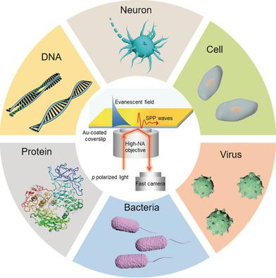 Figure 1. SPRM applications. (Zhou X L, et al., 2020).
Our Surface Plasmon Resonance Microscopy Testing
Creative Biostructure offers advanced SPRM testing to quickly and accurately detect interactions between biomolecules, providing strong technical support for research in the fields of drug discovery, biosensing, and disease diagnosis. Here is an overview of our SPRM workflow:
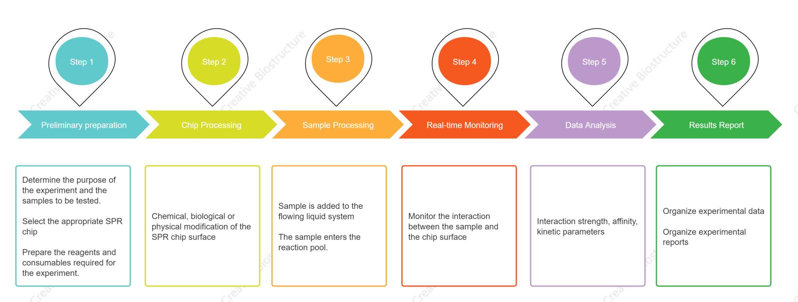 Figure 2. Our SPRM technology service process. (Creative Biostructure)
1. Preliminary Preparation: Identify the objectives and the specific biomolecular interactions to be studied. Choose the right SPR chip based on the requirements of the experiment.
2. Chip Processing: Modify the surface of the SPR chip to enhance its interaction with the sample. This may involve chemical treatments, biological coatings, or physical alterations.
3. Sample Processing: Gather all necessary materials and reagents for the experiment, ensuring that everything is ready for sample introduction.
4. Real-time Monitoring: Introduce the sample into the SPR system where it interacts with the chip surface in a controlled liquid environment. Utilize the SPRM technology to observe in real-time the interactions between the sample and the chip surface, capturing the binding kinetics and affinity.
5. Data Analysis: Collect and organize the data generated from the real-time monitoring, focusing on interaction strength, affinity, and kinetic parameters. Apply image reconstruction algorithms and resolution enhancement techniques such as azimuthal rotational illumination (ARI) to improve the quality of the SPRM images.
6. Results Report: Compile the findings into a comprehensive report detailing the biomolecular interactions observed, the strength of the interactions, and any relevant kinetic data. Ensure that the report is clear, concise, and presents the data in a manner that is accessible to the intended audience, whether it be scientific peers or stakeholders in a clinical or industrial setting.
With our professional team and cutting-edge equipment, we ensure that our customers can accurately interpret the complex relationships between biomolecules and promote rapid development and breakthroughs in their projects. If you are interested in our SPRM testing, please contact us for a detailed quote.
Key Advantages of Our SPRM Testing
- Precise Detection: Detects even minor changes in refractive index, enabling the capture of minute biomolecular interactions.
- High Resolution Imaging: Provides high-resolution imaging at the nanoscale, offering clear and detailed visualization of interaction sites.
- Dynamic Analysis: Allows for real-time observation of binding events, providing immediate insights into the interaction kinetics.
- Comprehensive Data: Offers both quantitative kinetic data (association and dissociation rates) and qualitative visual data for thorough analysis.
- Label-Free Technology: No need for fluorescent or radioactive labels, maintaining the native state of biomolecules.
- Expert Reporting: Comprehensive reports summarizing procedures, results, and insights, complemented by expert consultation.
Frequently Asked Questions
-
What types of samples are suitable for SPRM testing?
SPRM testing is suitable for a variety of sample types, including but not limited to biomolecules (proteins, nucleic acids, etc.), cell membranes, cell cultures, nanomaterials, and their surface modifications.
-
How does SPRM achieve nanometer-level resolution imaging?
SPRM utilizes the high sensitivity of surface plasmon waves to surpass the optical diffraction limit, achieving nanometer-level high-resolution imaging.
-
How are the results delivered and supported?
Upon completion, we provide a comprehensive report detailing all procedures, images, and quantified data. The report will include clear visuals and insights to facilitate your research. Additionally, our technical support team is available for consultations to help you interpret the results and provide further analysis if required. We ensure that you have all the support needed to make the most of your SPRM data.
Ordering Process

References
- Zhou X L, Yang Y, Wang S, et al. Surface plasmon resonance microscopy: from single-molecule sensing to single-cell imaging. Angewandte Chemie International Edition. 2020. 59(5): 1776-1785.
- Huang S, Chen J, Zhang T, et al. Recent advances in surface plasmon resonance microscopy. Chemosensors. 2022. 10(12): 509.
- Berthuy O I, Muldur S K, Rossi F, et al. Multiplex cell microarrays for high-throughput screening. Lab on a Chip. 2016. 16(22): 4248-4262.


