Application of Atomic Force Microscopy (AFM) in Materials Science
Atomic Force Microscope (AFM) is an analytical instrument that can study the surface structure of solid materials, including insulators. It studies the surface structure and properties of substances by detecting the extremely weak interatomic interaction between the surface of the sample to be tested and a miniature force-sensitive element. One end of a pair of microcantilever sensitive to weak force is fixed, and the tiny tip of the other end is close to the sample. It will interact with it at this time, and the force will make the microcantilever deform or change its motion state. When scanning the sample, use the sensor to detect these changes, and the force distribution information can be obtained to obtain the surface topography structure information and surface roughness information with nanometer resolution. AFM uses the physical phenomenon of the force interaction between the sample surface and the probe, so it is not limited by the requirement that the sample surface be conductive such as STM and can detect conductors. For insulators such as non-conductive tissues, biological materials, and organic materials, AFM can also obtain high-resolution surface topography images, which makes it more adaptable and has a broader application space.
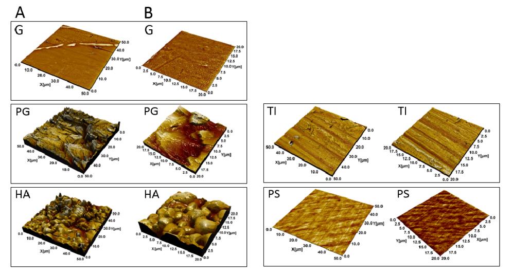
 Figure 1. Atomic Force Microscopy (AFM) topographical images of the materials.
Materials Science Applications of Our AFM Service
The materials science application of AFM provided by Creative Biostructure mainly includes these aspects: Topography observation, Composition Analysis, Nanomaterials and powder material analysis, and Crystal Growth Observation.
Topography observation
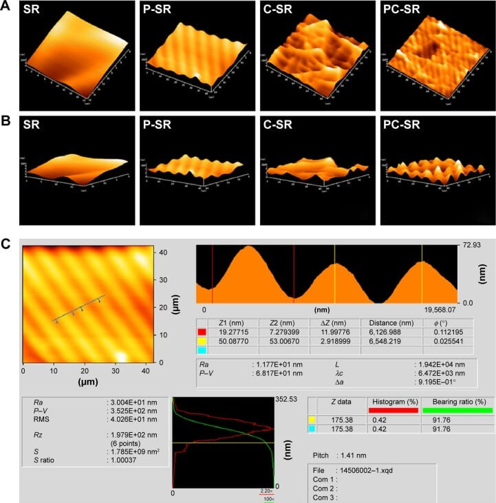
AFM has been widely used in various fields of surface analysis through the analysis, induction, and summary of surface topography to obtain deeper information. Since the ups and downs of the surface can be accurately obtained in numerical form, AFM can analyze the overall image of the surface to obtain parameters such as roughness, particle size, average gradient, pore structure, and pore size distribution of the sample surface; it can also perform a rich three-dimensional simulation display of the shape of the sample, making the image more suitable for intuitive observation.
Composition analysis
AFM can provide composition information according to the difference of some physical properties of materials in Phase Image mode.
Nanomaterials and powder material analysis
AFM is an effective analysis and testing tool for nanomaterials. The horizontal resolution of the atomic force microscope is 0.1-0.2nm, and the vertical resolution is 0.01nm, which can effectively characterize nanomaterials.
Crystal growth observation
Atomic force microscopy is a new and effective tool for atomic-level observation and study of crystal growth interface processes. Utilizing its high resolution and the ability to work in solution and atmospheric environments, it is possible to accurately observe the atomic-level resolution images of the growth interface in real-time and understand the growth process and mechanism.
 Figure 2. Surface topography of samples as observed by AFM.
Creative Biostructure can offer you the different applications of AFM in material samples, giving you the information you need to do great work. Please feel free to contact us for more information or a detailed quote.
Ordering Process
References
- Hiltunen A K., et al. Structural and functional dynamics of Staphylococcus aureus biofilms and biofilm matrix proteins on different clinical materials. Microorganisms. 2019, 7(12): 584.
- Lei Z., et al. Biofunctionalization of silicone rubber with microgroove-patterned surface and carbon-ion implantation to enhance biocompatibility and reduce capsule formation. International Journal of Nanomedicine. 2016: 5563-5572.
- Heath G R., et al. Localization atomic force microscopy. Nature. 2021, 594(7863): 385-390.
