Negative Staining TEM
Negative stain, also known as negative contrast stain, is a staining method commonly used for microscopic examination of opaque liquid specimens. Different from traditional dyeing, there is no reaction between the sample and the dye solution, so the sample is not colored. In contrast to the density difference between the sample and the dye, the white sample of low electron density can be contrasted with the black background, and then it will form an obvious negative contrast. As the sample does not need complex operations such as fixation, dehydration, embedding, and ultra-thin slicing, negative staining electron microscopy (Negative staining EM) has been widely used in the analysis of protein complex, viruses, bacteria, macromolecules, subcellular fragments, and so on. Creative Biostructure has extensive expertise and experience in negative staining EM technology.
Advantages of Negative staining EM
- Fasted speed and convenience.
- With lower voltage microscopes (such as 100 kV and 120 kV) requirements than higher voltage cryo-EM equivalents.
- Low demands of sample and dye solution.
- Using heavy metal stained to maximize the contrast to samples, and thus only fewer particles are required for data analysis.
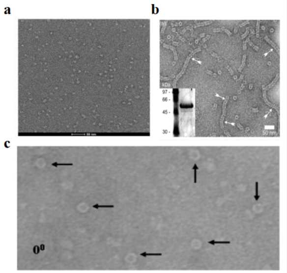 Figure 1. Negative stain of different samples
(a) Glucagon-like peptide-1 receptor with compound 2 complex[1];
(b) Recombinant Measles Virus[2];
(c) Neisseria meningitidis Secretin PilQ [3].
Our negative staining EM service covers the entire technical process, including sample preparation, EM data acquisition and analysis.
Sample preparation service
Preparing the sample of a suspension with a certain density firstly, then dispersing it on the copper net with supporting film, and finally dyeing with negative staining solution to increase the contrast, and then electron microscopic observation can be carried (Figure 2). Specifically, sample preparation can be divided into drop dyeing, flotation dyeing and spray dyeing.
There are many factors affecting the negative dyeing of samples, such as sample concentration, dye solution concentration and pH value, dyeing time and temperature, supporting membrane properties, dyeing type and so on. Creative Biostructure will choose the best condition to prepare samples.
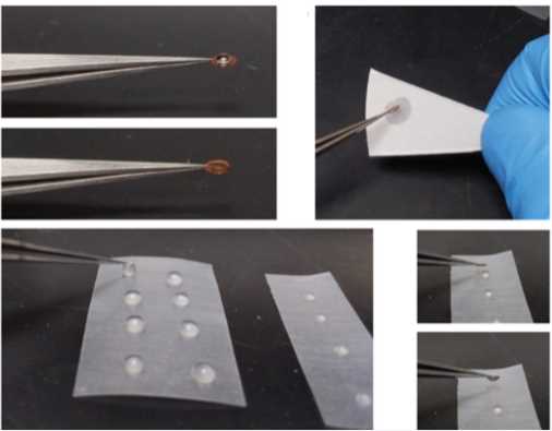 Figure 2. Process of negative staining sample preparation [4].
Data acquisition and analysis service
In basic, the homogeneity and dispersion of the prepared samples should be in good condition. A low-voltage (120 kV or 100 kV) transmission electron microscope is used for checking the quality of prepared samples. After obtaining good samples, sample images will be collected. The particles were pretreated and selected by using specific software such as RELION, and then by rounds of 2D average classification and optimization, the conformation of the target complex with low resolution would be obtained (Figure 3).
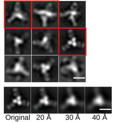 Figure 3. Conformation obtained by photo analysis collected by negative staining [4].
With our Negative staining EM platform, Creative Biostructure can provide sample preparation and data collection as well as analysis services for our customers. If you are interested in our negative-stain transmission electron microscopy service, please feel free to contact us. We are looking forward to cooperating with you.
Ordering Process

References
- Cong Z, et al. Molecular insights into ago-allosteric modulation of the human glucagon-like peptide-1 receptor. Nature Communications. 2021. 12(1): 1-11.
- Bhella D, et al. Conformational flexibility in recombinant measles virus nucleocapsids visualised by cryo-negative stain electron microscopy and real-space helical reconstruction. Journal of Molecular Biology. 2004. 340(2): 319-331.
- Collins R F, et al. Three-dimensional structure of the Neisseria meningitidis secretin PilQ determined from negative-stain transmission electron microscopy. Journal of Bacteriology. 2003. 185(8): 2611-2617.
- Gallagher J R, et al. Negative-stain transmission electron microscopy of molecular complexes for image analysis by 2D class averaging. Current Protocols in Microbiology. 2019. 54(1): e90.


