Exosome In Vitro Functional Assay
Exosomes are tiny vesicles secreted by the extracellular matrix with a diameter of 30-160 nm. Current studies have found that almost all types of cells can produce and release exosomes.
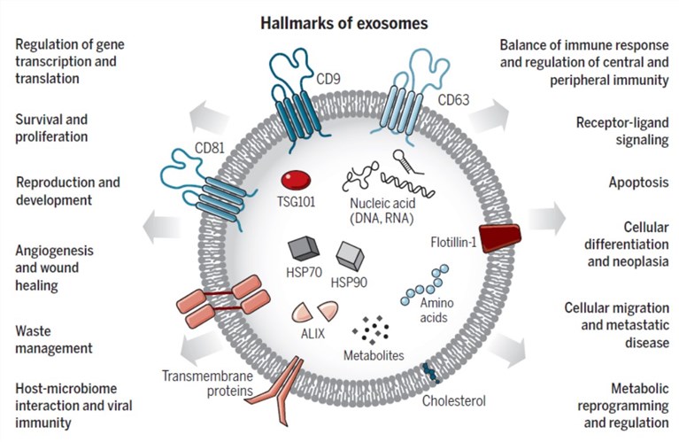 Figure 1. The main characteristics of exosomes. (LeBleu, et al. 2020)
With the molecular transfer function, exosomes may serve as a signal pathway for communication between cells. These exosomes are absorbed by recipient cells to communicate substances and signals through material exchange or release of inclusions.
We have a professional exosome research team with rich experience and focus on providing one-stop service. We can conduct in vitro analysis of the function of exosomes. For example, we can add labeled exosomes into the culture medium of recipient cells and co-culture with the recipient cells to observe the functional changes of the cells. For example, cell proliferation, migration and invasion, apoptosis, etc.
Commonly observed phenotypes after uptake of exosomes by recipient cells:
- CCK-8 cell proliferation assay
CCK-8 is a fast, highly sensitive, non-radioactive method based on WST-8 for cell proliferation and cytotoxicity detection. WST-8, a compound similar to MTT, can be reduced by some dehydrogenases in the mitochondria in the presence of electron-coupling reagents and produce orange-yellow crystals. The more and faster the cells multiply, the darker the color. Receptor cells in the logarithmic growth phase were inoculated into 96-well plates and treated with drugs (exosomes) after adhesion. Cell viability was detected by CCK-8 methods 24 h later.
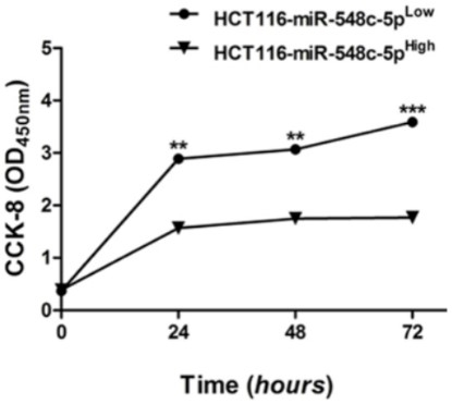 Figure 2. CCK-8 assay for the proliferation of HCT116 cells incubated with serum exosomes encapsulating cancer biomarker (miR-548c-5p). (Peng ZY, et al. 2018)
- Cell apoptosis detected by flow cytometry
The apoptosis or programmed cell death defines a gene-coded cell death program, which is distinct from necrosis or accidental cell death, in morphologically and biochemically. There are many techniques used to characterize apoptosis, of which flow cytometry is one of the most popular and widespread applications in the study of apoptosis. In the functional analysis of exosomes, the recipient cells of the logarithmic growth phase were inoculated into the orifice plate and treated with drugs (exosomes) after adhesion. After 48 hours, the cells were collected for staining and flow analysis.
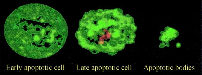 Figure 3. The apoptotic process. (Vermes I, et al. 2000)
- Cell cycle measured by flow cytometry
As the common application of flow cytometry application in Cell cycle analysis, with using a DNA-specific stain, the percentage of the population in G0/G1, S, and G2/M will be measured. In the exosome functional experiment, the logarithmic growth phase cells were inoculated into the petri dish and treated with drugs (exosomes) after attachment. After 24 hours, the cells were collected for fixed staining and flow analysis.
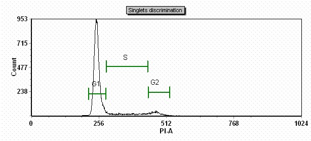 Figure 4. Cell cycle phase detection and analysis. (ROY J)
- Transwell assay for cell migration
Migration is a key property of live cells and critical for normal development, immune response, and disease processes such as cancer metastasis and inflammation. The effect of exosomes on the migration and invasion of cancer cells was determined by using Transwell 24-well plates.
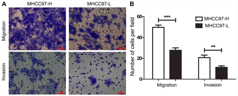 Figure 5. In vitro migration and invasion assays. (Wang S, et al. 2017)
- Cell invasion assay
Chamber on the kind of tumor cells, the room to join FBS or certain chemokines, tumor cells to the chamber to run under high nutrients, but unlike tumor cell migration experiment, polycarbonate membrane chamber side covered with a layer of matrix to mimic in vivo extracellular matrix, cell into the next room, to secrete matrix metalloproteinases (MMPs) matrix degradation, it can pass through the polycarbonate membrane. Counting the number of cells entering the inferior compartment can reflect the ability of tumor cells to invade.
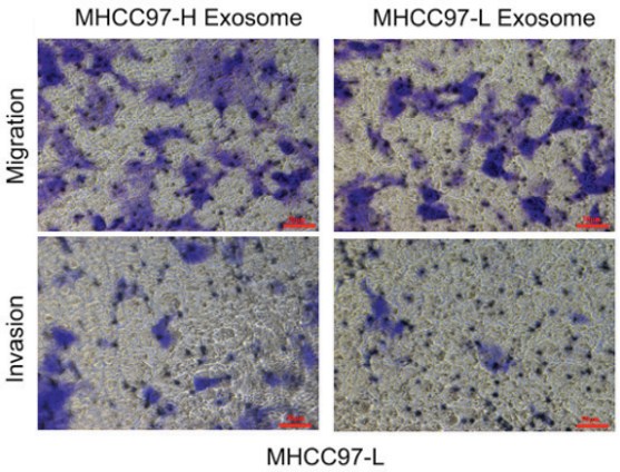 Figure 6. In vitro migration and invasion assays incubation with exosomes. (Wang S, et al. 2017)
Creative Biostructure offers different analysis for our customers, such as cell proliferation, apoptosis, cell cycle, cell migration/invasion, which helps to detect the cell function changes after treating by exosomes.
If you are interested in our in vitro functional assay service, please feel free to contact us. Creative Biostructure is looking forward to cooperating with you.
Ordering Process
References
- LeBleu, R.K.a.V.S., The biology, function, and biomedical applications of exosomes. Science. 2020, 367(6478).
- Peng ZY, et al. Downregulation of exosome-encapsulated miR-548c-5p is associated with poor prognosis in colorectal cancer. J Cell Biochem. 2018, 1-7.
- Vermes I, et al. Flow cytometry of apoptotic cell death. Journal of Immunological Methods. 2000, 243(1): 167-190.
- ROY J. CARVER BIOTECHNOLOGY CENTER https://biotech.illinois.edu/flowcytometry/protocols/cell-cycle-analysis
- Wang S, et al. Role of exosomes in hepatocellular carcinoma cell mobility alteration. Oncol Lett. 2017, 14(6): 8122-8131.
