Subcellular Organelle Structure
Subcellular organelles are specialized structures within cells that perform various functions critical to the survival and proper functioning of the cell. Understanding the structure and function of these organelles is fundamental to cell biology because each organelle contributes to the overall metabolic and physiological processes of the cell. The structural organization of these organelles varies greatly between prokaryotic and eukaryotic cells, reflecting the complexity and specialization required for the survival of different types of organisms.
- Prokaryotic vs. eukaryotic cells
- Nucleus
- Mitochondrion
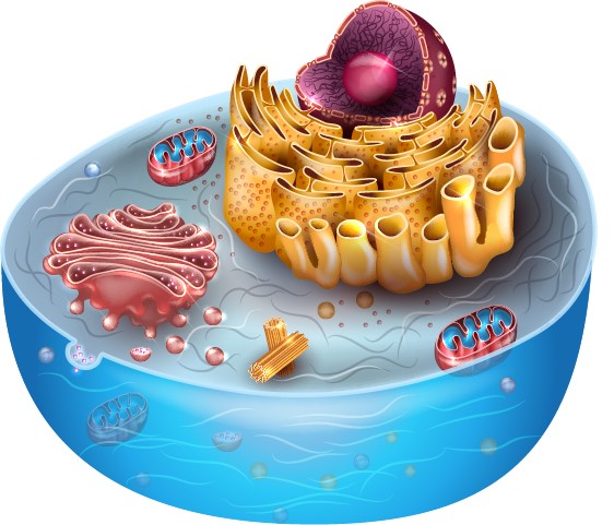
- Endoplasmic Reticulum (ER)
- Golgi Apparatus
- Lysosome and peroxisome
- Centrosome
- Chloroplast
- Ribosome
- Cytoskeleton
- Methods for studying organelle structure
Prokaryotic vs. Eukaryotic Cells
Cells can be broadly divided into two types: prokaryotic and eukaryotic. Prokaryotic cells, which include bacteria and archaea, are generally simpler and lack membrane-bound organelles. Instead, their cellular machinery is distributed throughout the cytoplasm. Prokaryotic cells have a nucleoid region where the genetic material is located, but they do not have a true nucleus. They rely on structures such as ribosomes for protein synthesis and may contain other specialized structures such as the cell wall, flagella, or pili, depending on their function and environment.
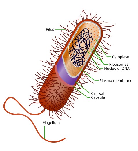 Figure 1. Diagram of a typical prokaryotic cell.
In contrast, eukaryotic cells, found in animals, plants, fungi, and protists, are characterized by their complexity and the presence of membrane-bound organelles. These cells have a true nucleus, which contains the genetic material of the cell and is surrounded by a nuclear envelope. Eukaryotic cells also contain a variety of organelles, each surrounded by membranes that compartmentalize cellular functions, allowing for greater specialization and efficiency. The presence of these organelles supports the more complex life processes associated with multicellular organisms.
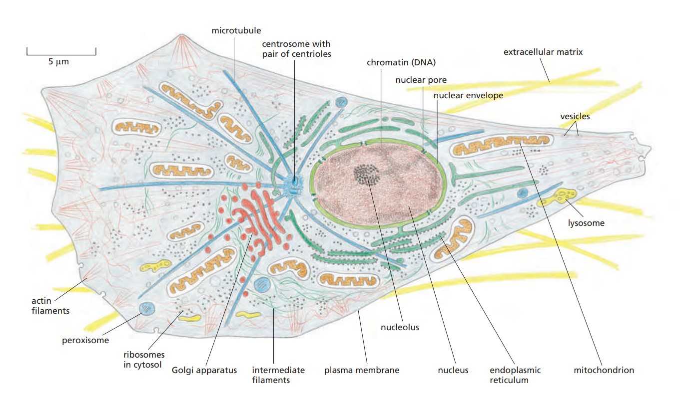 Figure 2. The major features of a typical animal cell (Molecular Biology of the Cell, 6th Edition).
Nucleus
The nucleus is the most prominent organelle in eukaryotic cells and serves as the control center of the cell. It is surrounded by a double membrane called the nuclear envelope, which is perforated with nuclear pores that regulate the exchange of materials between the nucleus and the cytoplasm. Inside the nucleus, chromatin (a complex of DNA and proteins) is organized into chromosomes during cell division. The nucleolus, a dense region within the nucleus, is the site of ribosomal RNA (rRNA) synthesis and ribosome assembly.
For more information about the structure of the nucleus, visit the following pages: Chromatin Structure, Nuclear Pore Complex Structure, and Nuclear Skeleton Structure.
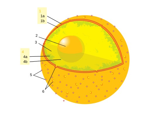 Figure 3. Structure of mammalian nucleus. 1: nuclear envelope; 1a: outer membrane; 1b: inner membrane; 2: nucleolus; 3: karyoplasm; 4: chromatin; 4a: heterochromatin; 4b: euchromatin; 5: ribosomes; 6: nuclear pores.
Mitochondrion
Mitochondria are often referred to as the powerhouses of the cell because of their role in generating ATP through oxidative phosphorylation. They are double-membrane organelles with a smooth outer membrane and an inner membrane that is highly folded into structures called cristae, which increase the surface area for energy production. Mitochondria contain their own DNA and ribosomes, reflecting their evolutionary origin from ancient symbiotic bacteria.
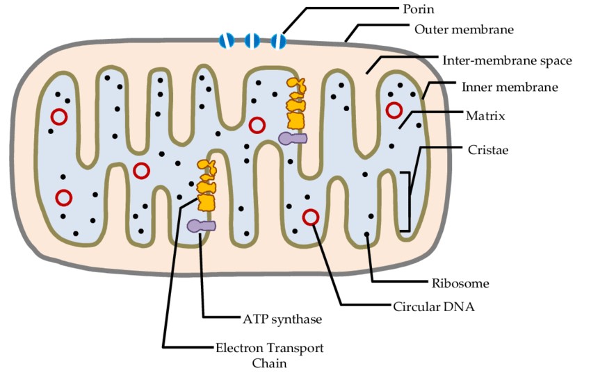 Figure 4. Structure of a mitochondrion (Yusoff et al., 2015).
Endoplasmic Reticulum (ER)
The ER is a network of membranous tubules and sacs that plays a central role in the synthesis of proteins and lipids. It exists in two forms: the rough ER, which is studded with ribosomes and is involved in protein synthesis and processing, and the smooth ER, which lacks ribosomes and is involved in lipid synthesis, detoxification, and calcium storage.
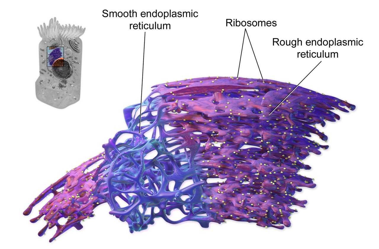 Figure 5. 3D rendering of endoplasmic reticulum.
Golgi Apparatus
The Golgi apparatus is a series of flattened membranous sacs that modify, sort, and package proteins and lipids for secretion or delivery to other organelles. It is often referred to as the "post office" of the cell because it is essential for processing and dispatching the molecular products of the ER.
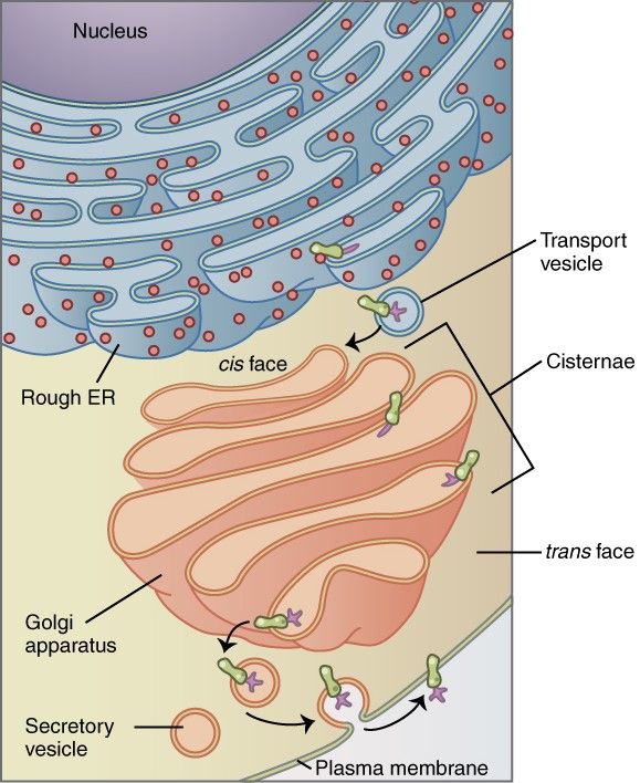 Figure 6. The Golgi apparatus (salmon pink) in context of the secretory pathway.
Lysosome and Peroxisome
Lysosomes are membrane-bound organelles that contain hydrolytic enzymes capable of breaking down various biomolecules, including proteins, nucleic acids, and lipids. They are crucial for intracellular digestion and recycling of cellular components. Acid hydrolases are active at the acidic pH maintained within the lysosome, but not at the neutral pH of the cytosol. The acidic internal pH of lysosomes results from the action of a proton pump in the lysosomal membrane that imports protons from the cytosol coupled to ATP hydrolysis.
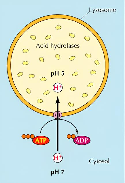 Figure 7. Organization of the lysosome (The Cell: A Molecular Approach, 2nd Edition).
Peroxisomes, on the other hand, are involved in the breakdown of fatty acids and the detoxification of harmful substances, such as hydrogen peroxide, which is converted to water and oxygen by the enzyme catalase.
Centrosome
The centrosome, also referred to as the central body, is a cell organelle with a diameter of approximately 1 µm. In most animal cells, it fulfills the function of a microtubule organizing center (MTOC) during the formation of the mitotic spindle, which is anchored at this location. The centrosome is typically situated in the center of the cell, in close proximity to the nucleus. It comprises a pair of cylindrical centrioles, oriented at right angles to one another, embedded in a protein matrix known as the pericentriolar matrix.
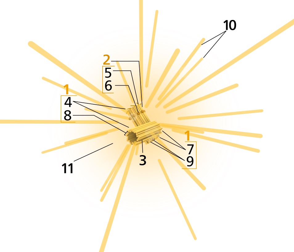 Figure 8. Components of a typical centrosome. 1: centriole; 2: mother centriole; 3: daughter centriole; 4: distal ends; 5: distal appendages; 6: subdistal appendages; 7: proximal ends; 8: microtubule triplets; 9: interconnecting fibers; 10: microtubules; 11: pericentriolar material.
Chloroplast
Chloroplasts are specialized organelles found in plant and algal cells and are responsible for the process of photosynthesis. They contain the pigment chlorophyll, which captures light energy and facilitates the conversion of carbon dioxide and water into glucose and oxygen. Similarly to mitochondria, chloroplasts possess their own DNA and are postulated to have arisen from symbiotic bacteria.
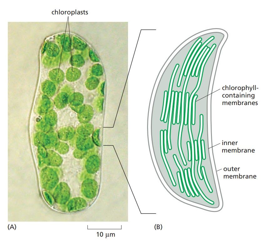 Figure 9. Chloroplasts. (A) A single cell isolated from a leaf of a flowering plant, seen in the light microscope, showing the green chloroplasts. (B) A drawing of one of the chloroplasts, showing the highly folded system of internal membranes containing the chlorophyll molecules by which light is absorbed (Molecular Biology of the Cell, 6th Edition).
Ribosome
Ribosomes are the molecular machines that carry out protein synthesis. They can be found free-floating in the cytoplasm or attached to the rough ER. Ribosomes are composed of rRNA and proteins and are divided into two subunits: the small subunit that reads the mRNA and the large subunit that assembles the amino acids into a polypeptide chain. Detailed information about the ribosome structure can be found here: Ribosome Structure.
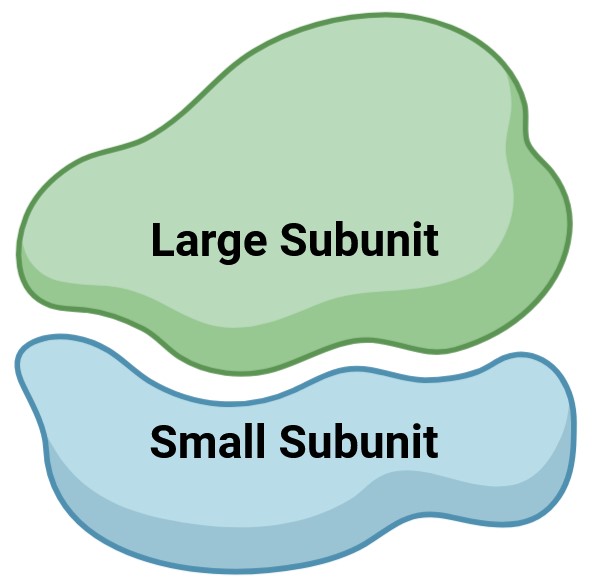 Figure 10. Large subunit and small subunit of ribosome (Created with BioRender.com).
Cytoskeleton
The cytoskeleton is a dynamic network of protein filaments that provides structural support to the cell, facilitates cell movement, and organizes organelles in the cytoplasm. It consists of three main types of filaments: microfilaments (actin filaments), intermediate filaments, and microtubules, each of which plays a different role in cell shape, transport, and division. To learn more about the structure of the cytoskeleton, click here: Cytoskeleton Structure.
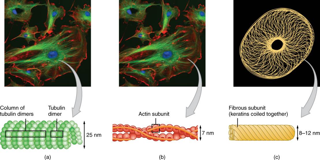 Figure 11. The cytoskeleton consists of (a) microtubules, (b) microfilaments, and (c) intermediate filaments.
Methods for Studying Subcellular Organelle Structure
The study of organelle structure has advanced significantly with the development of various imaging and biochemical techniques.
- Electron microscopy (EM), including transmission electron microscopy (TEM) and scanning electron microscopy (SEM), has been instrumental in revealing the detailed architecture of organelles at the nanometer scale. TEM provides detailed images of the internal structure of organelles, while SEM provides three-dimensional views of their surface. In addition, we have a professional team for cryo-EM structure analysis for subcellular organelle samples.
- Fluorescence microscopy, particularly confocal and super-resolution microscopy, has allowed scientists to visualize the dynamic behavior of organelles in living cells using fluorescent labels. These techniques allow the study of organelle interactions, movements, and changes in real time.
- Biochemical methods, such as subcellular fractionation and mass spectrometry are used to isolate and analyze the molecular composition of organelles. These techniques help identify the specific proteins, lipids, and other molecules associated with each organelle and provide insight into their function.
- Molecular biology techniques, including gene editing (e.g., CRISPR-Cas9), allow researchers to manipulate genes encoding organelle components and to study the effects of these modifications on organelle structure and function.
In summary, subcellular organelles are complex structures that play specialized roles in the life of a cell. The distinction between prokaryotic and eukaryotic cells underscores the evolutionary complexity that has led to the diversity of life. Advances in imaging and biochemical techniques have provided detailed insights into the structure and function of these organelles, with far-reaching implications for medicine, biotechnology, and basic biology.
Creative Biostructure is dedicated to the detailed analysis of subcellular structures. Understanding the structure of organelles not only deepens our knowledge of cellular processes, but also offers potential for innovation in various scientific and industrial fields. Our expertise sheds light on the structural organization and functional roles of these tiny components, providing invaluable insights for medicine, biotechnology, and basic biology. For more information on how our services can support your research, please contact us for a customized quote.
References
- Molecular Biology of the Cell, 6th Edition.
- The Cell: A Molecular Approach, 2nd Edition.
- Yusoff, A. A. M., Ahmad, F., Idris, Z., Jaafar, H., & Abdullah, J. M. (2015). Understanding mitochondrial DNA in brain tumorigenesis. In Molecular Considerations and Evolving Surgical Management Issues in the Treatment of Patients with a Brain Tumor.

