How to Track Exosomes?
Exosomes are nanosized lipid bilayer vesicles released by almost all cells and are involved in intercellular communication and transfer of substances. In recent years, exosomes have been recognized as biomarkers for diagnosing diseases and monitoring therapeutic effects. In addition, exosomes are expected to be used as natural carriers for therapeutic agents and drug delivery. To further advance the research and application of exosomes, an accurate understanding of the specific metabolic pathways in vitro and in vivo is required. Exosome tracking can help researchers understand the uptake mechanisms, biodistribution, migration, and therapeutic properties.
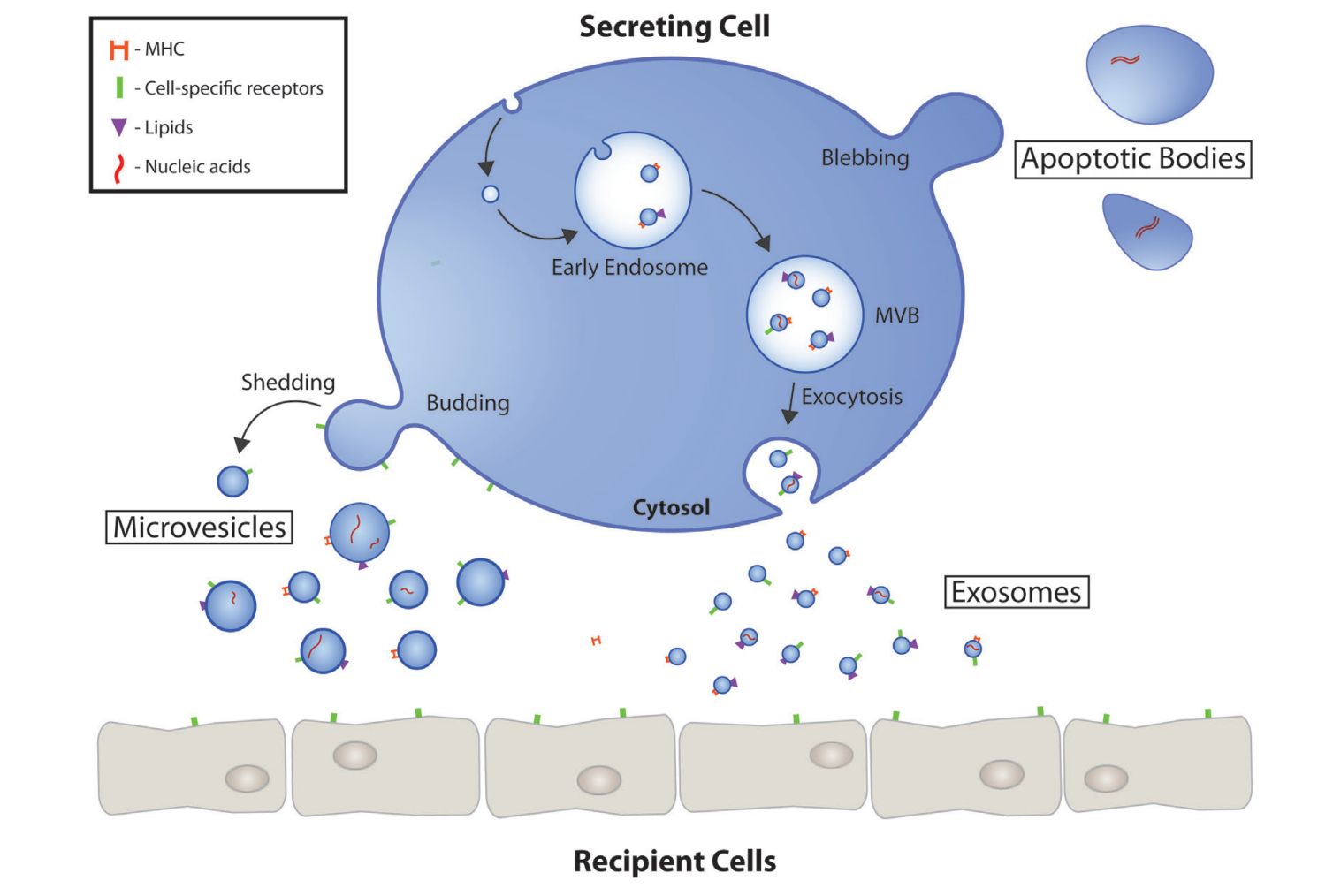 Figure 1. Extracellular vesicle (EV) biogenesis and secretion. (Gustafson D, et al., 2017)
What is Exosome Tracking?
Exosomes are small vesicles about 30-150nm secreted by living cells with a typical lipid bilayer structure that are involved in cell-to-cell communication, transport of substances, and maintenance of normal physiological processes, in addition to being implicated in disease production. Exosome tracking is the process of monitoring and visualizing the movement, biodistribution, and fate of exosomes within a biological system. It involves the use of labeling technologies and imaging methods to track the dynamics and behavior of exosomes. Labeling exosomes by tracers (e.g., organic dyes, fluorescent proteins, contrast agents, and nuclides.) and tracking of exosomes in animals using specific imaging methods allow observation of the biological behavior in vivo and detection of endocytosis of exosomes by target cells, which contribute to further research on exosome function.
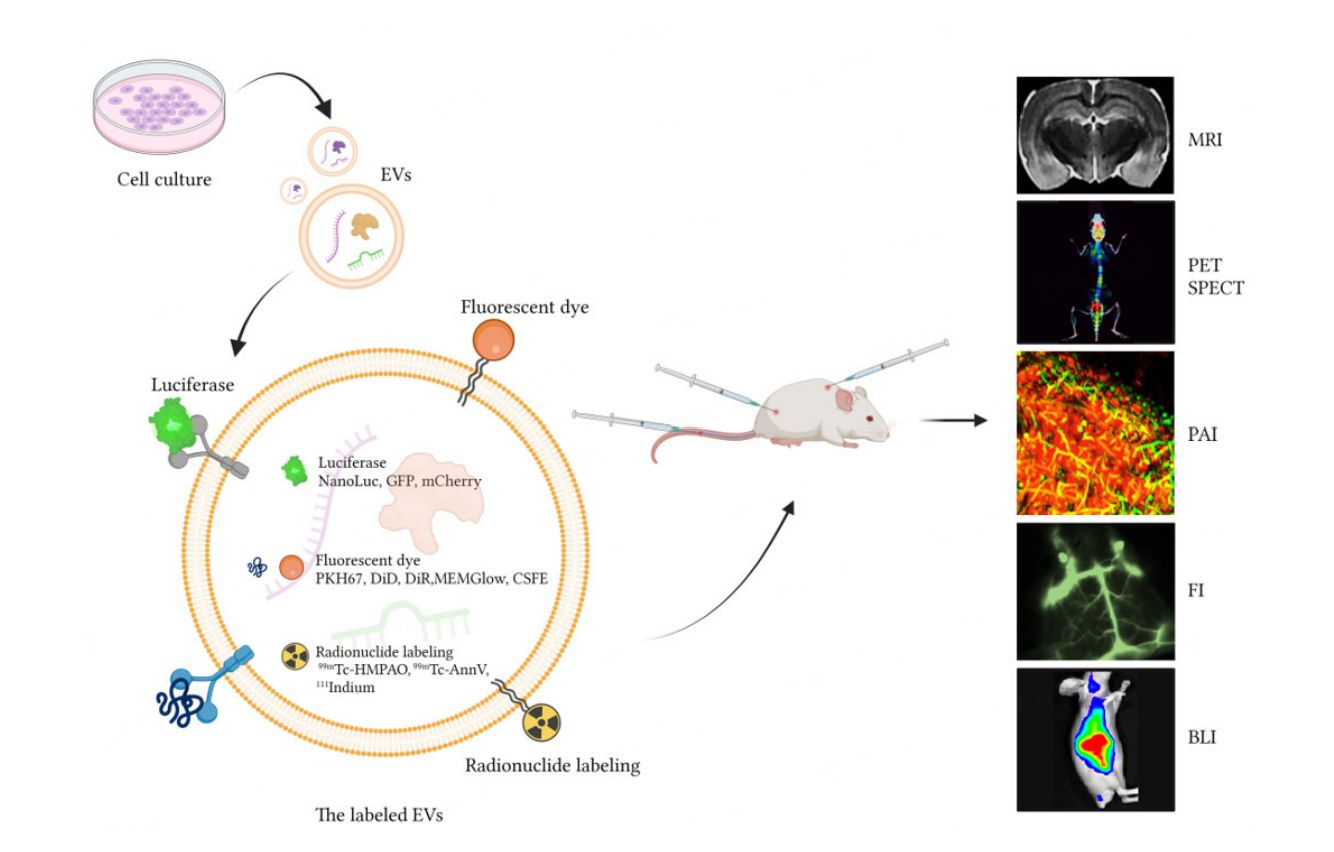 Figure 2. Strategies for tracking extracellular vesicles in vivo. (Shi Y, et al., 2023)
Tracking is an essential step in the field of exosome research, allowing researchers to gain insight into multiple aspects of exosome biology:
- Biodistribution and tissue targeting.
- Uptake and internalization.
- Transport and intracellular dynamics.
- Clearance and elimination.
- Heterogeneity and subpopulations.
Exosome Tracking Technologies
| Technologies | Tracer element | Advantages | Limitations |
| Fluorescence Imaging | DiR, Cy5.5, enhanced GFP, AIEgens, and pHluorin | High sensitivity, real-time imaging, versatile labeling options, and visualization of subcellular localization. | Limited tissue penetration, potential phototoxicity, and background fluorescence from unlabeled components. |
| Bioluminescence Imaging | RLuc, Gaussian luciferase, NanoLuc, and ThermoLuc | High sensitivity, low background, no external excitation light required. | Requires genetic modification of exosomes, limited tissue penetration, and low spatial resolution. |
| Nuclear Imaging | 99mTc, 111Indium, and 64Cu | Deep tissue penetration, high sensitivity, and ability to quantify exosome biodistribution. | Exposure to ionizing radiation, complex radiolabeling procedures, and potential alterations in exosome properties. |
| Magnetic Resonance Imaging | SPIONs, gadolinium, GIONs, and FTH1 | High spatial resolution, excellent soft tissue contrast, and no ionizing radiation. | Lower sensitivity compared to nuclear imaging, potential interference from magnetic nanoparticle labeling, and longer imaging times. |
| Photoacoustic Imaging | Ce6-R, DiR, Au Nanostars and TDSP | Deep tissue penetration, high spatial resolution, label-free detection of endogenous exosomal components. | The ability to quantify exosome concentrations is limited. |
| Computed Tomography | Glu-GNPs | High spatial resolution enables visualization of anatomical structures. | Exposure to ionizing radiation, limited soft tissue contrast, and low sensitivity for exosome detection. |
What are the Applications of Exosome Tracking?
Drug delivery and therapeutic monitoring
- Assess delivery and accumulation of exosome-encapsulated drugs or therapeutic agents.
- Monitor pharmacokinetics and pharmacodynamics of exosome-based therapies.
- Optimize exosome-mediated drug delivery strategies.
Diagnostics and Biomarker Development
- Track the release and clearance of disease-associated exosomes to identify potential biomarkers.
- Monitor changes in exosome profiles as a diagnostic tool for various diseases.
- Evaluate the utility of exosomes as markers for non-invasive liquid biopsies.
Preclinical research
- Assess the in vivo fate and efficacy of exosome-based therapies in animal models.
- Monitor exosome distribution and clearance in preclinical trials.
- Assess the safety and potential adverse effects of exosome-based interventions.
By utilizing diverse labeling methods and imaging techniques to track exosomes, Creative Biostructure can support a wide range of exosome research applications. In addition, with cutting-edge equipment and specialized knowledge in exosome research, we provide one-stop exosome services, including exosome isolation, characterization, labeling, tracking, targeting, and functional analysis, to help clients develop innovative exosome-based therapies. If you are interested in our services, please feel free to contact us for a formal quotation.
References
- Gustafson D, et al. Extracellular Vesicles as Protagonists of Diabetic Cardiovascular Pathology. Front Cardiovasc Med. 2017. 4: 71.
- Shi Y, et al. Research progress in in vivo tracing technology for extracellular vesicles. Extracellular Vesicles and Circulating Nucleic Acids. 2023. 4(4): 684-97.
- Liu Q, et al. Tracking tools of extracellular vesicles for biomedical research. Front Bioeng Biotechnol. 2022. 10: 943712.
- Jiang A, et al. In Vivo Imaging for the Visualization of Extracellular Vesicle-Based Tumor Therapy. ChemistryOpen. 2022. 11(9): e202200124.


