How to Characterize Exosomes?
The communicative properties of exosomes play an important role in the development of many diseases and are considered promising biomarkers for the diagnosis and prognosis of various diseases. In addition, exosomes can encapsulate cargo, cross biological barriers, and be biocompatible, making them an attractive prospect for next-generation therapies. This has led to a growing interest in the study of exosomes.
Exosome characterization offers tremendous clinical opportunities in the field of liquid biopsy. This potential requires high precision and reproducibility in the detection and characterization of exosomes. Creative Biostructure offers clients a full range of experience and technical capabilities to facilitate the comprehensive analysis of exosomes.
What are Exosomes?
Exosomes are a specific subclass of extracellular vesicles that range in size from 30 to 150 nm and can be found in a variety of body fluids, including plasma, cerebrospinal fluid, and saliva. Exosomes are packed with biological materials such as small RNAs, mRNAs, bioactive lipids, and proteins to convey critical information. As key mediators of intercellular communication, exosomes play important roles in physiology and disease development including tissue homeostasis and development. Two of the best-known examples are in cancer, where exosomes promote metastasis, angiogenesis, and chemotherapy resistance; and in neurodegenerative diseases such as Parkinson's disease, where exosomes shuttle toxic α-synuclein between cells and induce apoptosis.
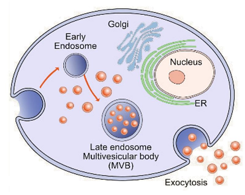 Figure 1. Schematic representation of exosome. (Gurunathan S, et al., 2019)
Why Exosome Characterization?
Exosomes are currently used in clinical trials for a wide range of diseases as direct therapeutic mediators or biomarkers of therapeutic response. The quality and purity of isolated exosomes are critical for their functional studies and may directly influence experimental results. To improve therapeutic efficacy to provide key insights and in-depth assessment of their mode of action, to better define exosome characteristics, and to facilitate downstream applications, it is essential to isolate and characterize exosomes using appropriate methods. To fully understand the biology and interactions of exosomes, a comprehensive understanding of their physicochemical properties, including size, shape, density, surface charge, and porosity, is essential.
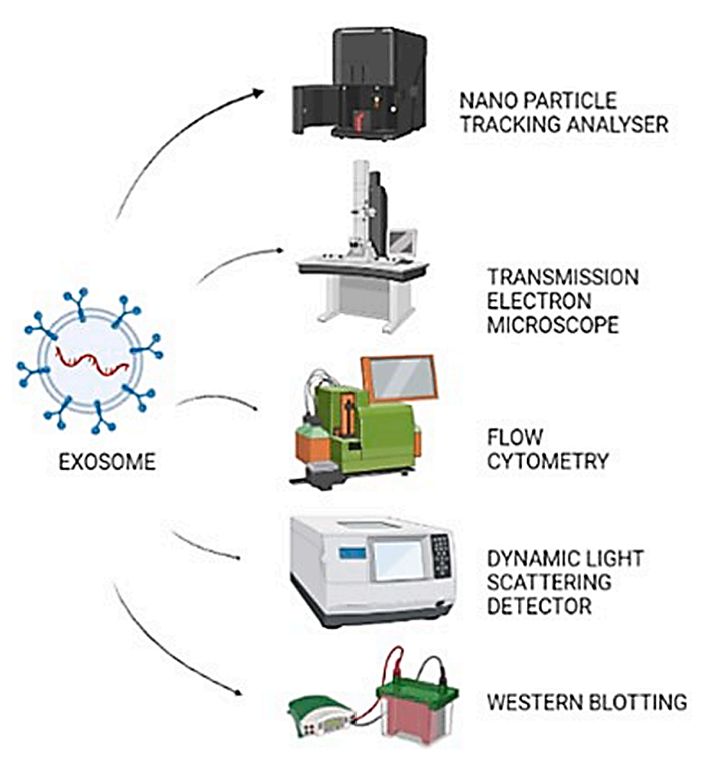 Figure 2. Common characterization techniques of exosomes. (Panigrahi AR, et al., 2022)
Our Exosome Characterization Technologies
| Technologies | Description |
| Transmission Electron Microscopy (TEM) | TEM is a technique widely used to characterize the structure, morphology, and size of a variety of biological components.TEM is specifically designed for visualization and the images obtained can be used for exosome diameter measurements. |
| Nanoparticle Tracking Analysis (NTA) | The NTA technique can characterize exosomes in the 10nm-2µm and the output is particle size, size distribution, concentration, and phenotype. Sample preparation is very fast and easy using this method and measurements take only a few minutes. |
| Dynamic Light Scattering (DLS) | The DLS technique is capable of measuring particles from 1nm to 6µm and is best suited for measuring one type of particle in suspension (monodisperse suspensions). This method is simple to use but does not allow visualization of the particles. |
| Atomic Force Microscope (AFM) | AFM is used as a nanoscale tool to characterize the abundance, morphology, biomechanics, and biomolecular composition of exosomes. It helps to study exosomes at the single vesicle and subvesicle level. It is capable of measuring samples under natural conditions and is a non-destructive mode of operation. |
| Flow Cytometry | Flow cytometry is a molecular method used to characterize exosome surface proteins, which can also measure the exosome size and structure. It can analyze different physical and chemical characteristics of cells and particles in suspension and can quantify or classify exosomes based on antigen expression levels. |
| Tunable Resistive Pulse Sensing (TRPS) | TRPS is a new technique to measure the size distribution and concentration of exosomes and to characterize colloidal particles ranging from ~50 nm to cell size. The feature of this technique is the in situ single particle characterization and concentration measurement of exosomes. |
| Western Blot | The technique is well established and allows qualitative and quantitative analysis of labeled proteins. It is also easier to analyze exosomes from cell culture media. However, it is not suitable for detecting exosomal labeled proteins in biological fluids. |
| ELISA | The method is highly specific, fast, and can qualitatively and quantitatively analyze exosome-tagged proteins, making it suitable for high-throughput analysis. However, the measurement results have poor reproducibility and many interfering factors. |
Related Products
Quantifying and characterizing exosomes is not an easy task. Their small size, low protein content, and co-purification with lipoproteins pose significant challenges for most analytical platforms. To better understand exosome properties and facilitate later therapeutic and diagnostic applications, we offer exosome isolation kits and a comprehensive range of exosome products. Our highly purified exosome products can be used as positive controls or directly for functional studies.
| Cat No. | Product Name | Source |
| Exo-UEK01 | Urine Exosome Kit | / |
| Exo-HDBF-01 | HQExo™ Exosome-SDH-Alzheimer's plasma | Exosome derived from Single Donor Human Alzheimer's plasma |
| Exo-HDBF-02 | HQExo™ Exosome-SDH-Asthma plasma | Exosome derived from Single Donor Human Asthma plasma |
| Exo-EV-A-001 | HQExo™ Exosome-Bovine Plasma exosome | Exosome derived from Bovine Plasma |
| Exo-EV-A-002 | HQExo™ Exosome-Canine Beagle Plasma exosome | Exosome derived from Canine Beagle Plasma |
| Exo-BF01 | HQExo™ Exosome-CSF | Exosome derived from human pooled CSF (healthy donors) |
| Exo-BF12 | HQExo™ Exosome-Human biofluids ascites fluid | Exosome derived from Human Ascites Fluid (healthy donors) |
| Exo-CH08 | HQExo™ Exosome-BPH-1 | Exosome derived from human benign prostatic hyperplasia-1 (BPH-1 cell line) |
| Exo-CH23 | HQExo™ Exosome-BxPC-3 | Exosome derived from human pancreas carcinoma cell line (BxPC-3 cell line) |
| Exo-CM01 | HQExo™ Exosome-B16F10 | Exosome derived from murine melanoma cell line (B16F10 cell line) |
| Exo-CH15 | HQExo™ Exosome-BLCL21 | Exosome derived from EBV transformed lymphoblastoid B cells (BLCL21 cell line) |
| Explore All Exosome Products | ||
For exosome studies to be reliable, reproducible, and rigorous, it is important to fully characterize exosomes. At Creative Biostructure, we have a range of advanced technologies and comprehensive products to help clients achieve this goal. Contact us for a wide range of exosome products and services to advance your research.
References
- Gurunathan S, et al. Review of the Isolation, Characterization, Biological Function, and Multifarious Therapeutic Approaches of Exosomes. Cells. 2019. 8(4): 307.
- Panigrahi AR, et al. Exosomes: Insights and therapeutic applications in cancer. Transl Oncol. 2022. 21: 101439.
- Lai JJ, et al. Exosome Processing and Characterization Approaches for Research and Technology Development. Adv Sci (Weinh). 2022. 9(15): e2103222.
- Zhang Y, et al. Exosome: A Review of Its Classification, Isolation Techniques, Storage, Diagnostic and Targeted Therapy Applications. Int J Nanomedicine. 2020. 15: 6917-6934.


