How to Label Exosomes?
Exosomes are produced by all known eukaryotic cells and constitute an essential means of intercellular communication. Recent research has revealed the essential role of exosomes in migration to specialized sites and cells. Functional research in in vivo and in vitro systems first requires exosome labeling.
Overview of Exosomes
Exosomes are particles encapsulated by lipid bilayers ranging from 30 to 150 nm and contain a series of membrane-associated higher-order oligomeric protein complexes that display significant molecular heterogeneity. Exosomes carry specific proteins, lipids, nucleic acids, and glycan affixes and are part of intercellular signaling networks in multicellular tissues. They mediate a wide range of activities and play essential roles in many aspects of human health and disease, including development, immunity, tissue homeostasis, cancer, and neurodegenerative diseases. Due to the known physiological and pathological functions, the clinical application of exosomes as biomarkers, therapeutic agents, and molecular carriers of therapeutics has been recognized as promising with remarkable results.
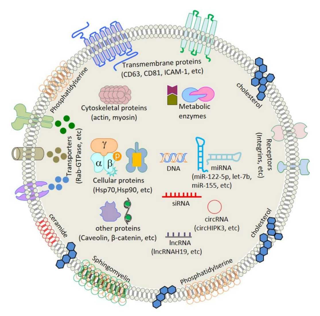 Figure 1. General representation of the exosome structure. (Gaurav I, et al., 2021)
The formation, release, recycling, uptake, and content delivery of exosomes have been critical in cancer diagnostic and therapeutic research. The translation and application of the research results are still in their infancy, and adequate exosome labeling and tracking techniques are needed to help researchers study the biological properties of exosomes in depth.
What is Exosome Labeling and How Does It Facilitate Research?
Exosome labeling involves attaching markers or tags to the surface or interior of an exosome to track its movement, uptake, and research its interactions with other substances in the environment. This process allows researchers to observe and research exosomes more efficiently and visually. By labeling exosomes, scientists can better understand their behavior, distribution, and function in biological systems. This approach is essential in many areas of research, including understanding disease mechanisms, developing diagnostic tools, and researching potential therapeutic applications of exosomes. Labeling can enhance understanding of exosome-mediated cellular communication and plays a crucial role in advancing the understanding of exosome biology and its role in health and disease.
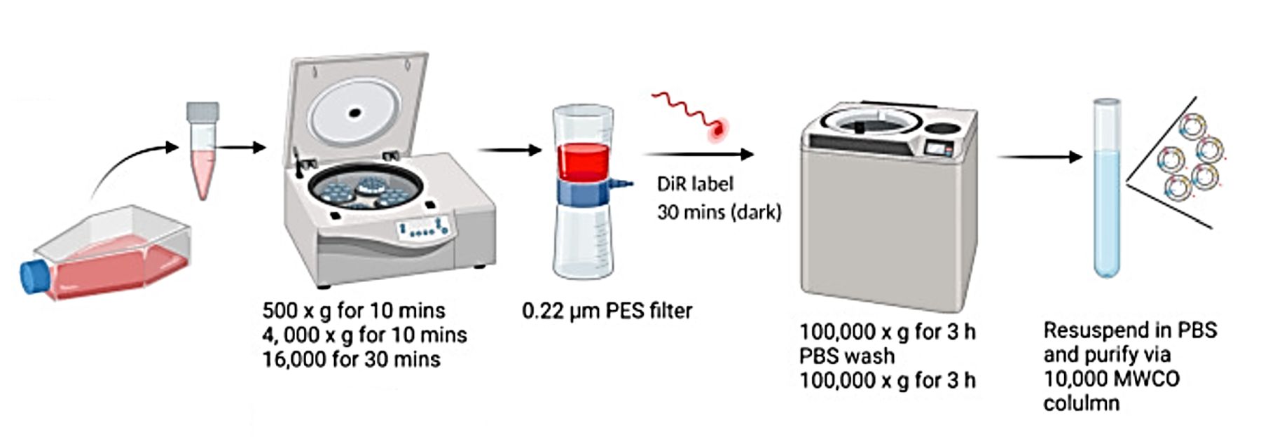 Figure 2. The EV purification and labeling scheme. (Gaurav I, et al., 2021)
What are the Commonly Used Techniques for Exosome Labeling?
| Techniques | Description | Advantages | Limitations |
| Lipophilic Dyes | Lipophilic dyes, such as PKH26 or DiO, penetrate the exosome membrane and produce fluorescence. | Lipophilic dyes are relatively simple to use, fluoresce brightly and stably, and are compatible with a variety of downstream applications, such as fluorescence microscopy and flow cytometry. | Over time, lipophilic dyes may lead to non-specific labeling. In addition, some lipophilic dyes may exhibit cytotoxicity or affect exosome behavior. |
| Lentivirus-Mediated CD63-GFP Expression | The introduction of the GFP gene into cells using genetic modification produces exosomes that carry the GFP protein and can be detected fluorescently. | Lentiviral-mediated expression ensures stable and heritable markers that enable long-term tracking of exosomes and improve specificity. | Genetic modification of cells alters the properties of exosomes and may affect their biological functions. |
| Gold Nanoparticles (GNPs) | Gold nanoparticles functionalized with specific ligands are attached to exosome membranes for visualization and tracking. | Gold nanoparticles provide strong and stable signals that can be detected using electron microscopy and spectroscopy due to their unique optical properties. | The size and surface properties of gold nanoparticles may affect exosome behavior and interactions. |
| Near Infrared (NIR) Fluorescent Probes | Near-infrared fluorophores are used to label exosomes, and the NIR probe emits fluorescence in the near-infrared range to penetrate deep tissues and reduce background autofluorescence. | NIR probes enable non-invasive imaging and tracking of exosomes in vivo with high sensitivity and specificity. | The availability of specific NIR probes for exosome labeling may be limited. |
| Radioisotopes | Radioisotopes are integrated into the exosome membrane, enabling detection using radioisotope imaging. | Sensitive, quantitative detection of exosomes with excellent tissue penetration. | Handling of radioactive materials requires specialized facilities and expertise. Radioisotopes may have potential cytotoxic effects on exosomes and receptor cells. |
| Membrane-permeable Compound Tracers | Fluorescent dyes or tracers permeable to membranes diffuse into exosomes and emit fluorescence. | Membrane permeable dyes are easy to use and provide uniform labeling of exosomes for real-time tracking. | Similar to lipophilic dyes, leakage of dyes from exosomes over time may result in nonspecific labeling and potential interference with quantitation. |
Our Services and Products
As a leading biological company providing labeling exosome service, Creative Biostructure can select appropriate labeling methods based on labeling efficiency, stability, compatibility with downstream applications, and clients' requirements.
We can label exosomes from a variety of sources, including cell culture supernatants, biological fluids, and tissue samples. Samples are required to contain adequate exosome yield, purity, and viability. We also offer exosome isolation services and high-quality exosome products for clients to choose from.
| Cat No. | Product Name | Source |
| Exo-CH04 | HQExo™ Exosome-H196 | Exosome derived from Human small cell lung cancer cell line (H196 cell line) |
| Exo-CH05 | HQExo™ Exosome-H841 | Exosome derived from human small cell lung cancer cell line (H841 cell line) |
| Exo-IC06 | HQExo™ Exosome-NK | Exosome derived from human NK cell line |
| Exo-SC03 | HQExo™ Exosome-hTERT | Exosome derived from hTERT-immortalized Mesenchymal Stem Cell |
| Exo-SC04 | HQExo™ Exosome-MSC | Exosome derived from Xeno-Free Human Mesenchymal Stem/Stromal Cells and Media |
| Exo-HDBF-03 | HQExo™ Exosome-SDH-Atopic Dermatitis plasma | Exosome derived from Single Donor Human Atopic Dermatitis plasma |
| Exo-HDBF-04 | HQExo™ Exosome-SDH-Benign Breast Conditions plasma | Exosome derived from Single Donor Human Benign Breast Conditions plasma |
| Explore All Exosomes Products | ||
Creative Biostructure provides efficient exosome labeling and tracking services. You can contact us with your requirements based on your research objectives and we will develop the most appropriate plan for you.
References
- Jan AT, et al. Expedition into Exosome Biology: A Perspective of Progress from Discovery to Therapeutic Development. Cancers (Basel). 2021. 13(5): 1157.
- Santelices J, et al. Fluorescent Labeling of Small Extracellular Vesicles (EVs) Isolated from Conditioned Media. Bio Protoc. 2022. 12(12): e4447.
- Bao C, et al. A Review of Labeling Approaches Used in Small Extracellular Vesicles Tracing and Imaging. Int J Nanomedicine. 2023. 18: 4567-4588.

