Exosome Isolation by Immunoaffinity (IA)
Exosomes play an irreplaceable role in early detection and therapy, but their small size and mixing with similar components pose a great challenge for their isolation. The correct elucidation of exosome-mediated physiological and pathological processes largely depends on the reliability of isolation and purification methods. Immunoaffinity-based isolation methods can represent an effective solution to obtain high-purity exosomes.
What is Immunoaffinity (IA)?
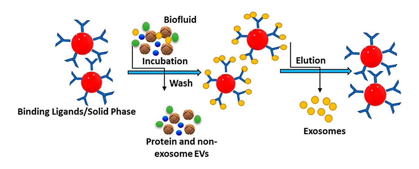
IA is a technique for isolating and purifying biological particles based on antigen-antibody-specific reactions. The membranes of exosomes are rich in proteins and receptors. These ubiquitous proteins (e.g., transmembrane and membrane-bound proteins) can bind to the corresponding antibodies and isolate exosomes.
 Figure 1. The principle of immunoaffinity-based method for isolating exosomes. (Sonbhadra S, et al., 2023)
In addition, by targeting specific proteins contained in exosomes, effective isolation of cancer-derived exosomes can be achieved. Generally, antibodies need to be immobilized on carriers, such as magnetic particles, chromatographic substrates, and microfluidic devices. Currently, the more common ones are immunomagnetic beads. Antibody-coated beads specifically bind the corresponding exosomes, distinguishing them from unbound impurities through magnetism.
How to Isolate Exosomes by IA Technology?
Exosome isolation by IA technology involves the use of specific antibodies or affinity capture agents to selectively capture exosomes based on their surface markers or antigens. Here is a description of the process:

1. Antibody or affinity selection - The first step in IA-based exosome isolation is the selection of an antibody or affinity capture agent against a specific exosome marker or antigen.
2. Coating of capture beads or surfaces - Immobilization of selected antibodies or capture agents onto solid support materials such as magnetic beads or microtiter plates.
3. Sample incubation- The sample-containing sample is incubated with the antibody-coated capture beads or surface. Exosomes present in the sample bind specifically to the immobilized antibody or capture agent via their surface markers.
4. Wash - After incubation, unbound components, including non-exosome particles and contaminants, are removed through a series of washing steps to ensure the purification of exosomes.
5. Elution - The specifically captured exosomes are eluted from the antibody-coated capture surface or beads. Elution can be achieved using a variety of methods, such as changing pH, using a mild elution buffer, or employing enzymatic digestion.
6. Post-isolation processing - After elution, the isolated exosomes can undergo further processing steps, such as concentration, purification, or characterization.
Application of IA Technology to the Isolation of Cancer-Derived Exosomes
IA technology is being used as a powerful tool for the isolation of cancer-derived exosomes. This method takes advantage of the presence of specific biomarkers on the surface of exosomes originating from cancer cells. By using antibodies directed against these biomarkers, IA technology can selectively capture and isolate cancer-derived exosomes from complex biological samples. This allows for the study and characterization of exosomal content, which provides insights into cancer pathology and can potentially be used as diagnostics markers or therapeutic targets. In addition, these exosomes can also be utilized for drug delivery in cancer treatment.
If you're exploring the role of exosomes in cancer diagnosis and treatment, take a look at Creative Biostructure's offerings. We are a leading supplier of high-purity exosome products, and our cancer cell-derived exosome products can be used directly for biomarker discovery and functional characterization, as well as providing insight into anti-tumor research.
| Cat No. | Product Name | Source |
| Exo-CH21 | HQExo™ Exosome-A375 | Exosome derived from human malignant melanoma cell line (A375 cell line) |
| Exo-CH07 | HQExo™ Exosome-MDA-MB-231 | Exosome derived from human breast cancer, aggressive/invasive/metastatic cell line (MDA-MB-231 cell line) |
| Exo-CH15 | HQExo™ Exosome-BLCL21 | Exosome derived from EBV transformed lymphoblastoid B cells (BLCL21 cell line) |
| Exo-CH10 | HQExo™ Exosome-HCT116 | Exosome derived from human colorectal carcinoma cell line (HCT116 cell line) |
| Exo-CH08 | HQExo™ Exosome-BPH-1 | Exosome derived from human benign prostatic hyperplasia-1 (BPH-1 cell line) |
| Exo-CH14 | HQExo™ Exosome-SK-N-SH | Exosome derived from human neuroblastoma (SK-N-SH cell line) |
| Explore All Exosomes Isolated from Cancer Cell Lines | ||
New Insights into Capturing Exosomes by IA
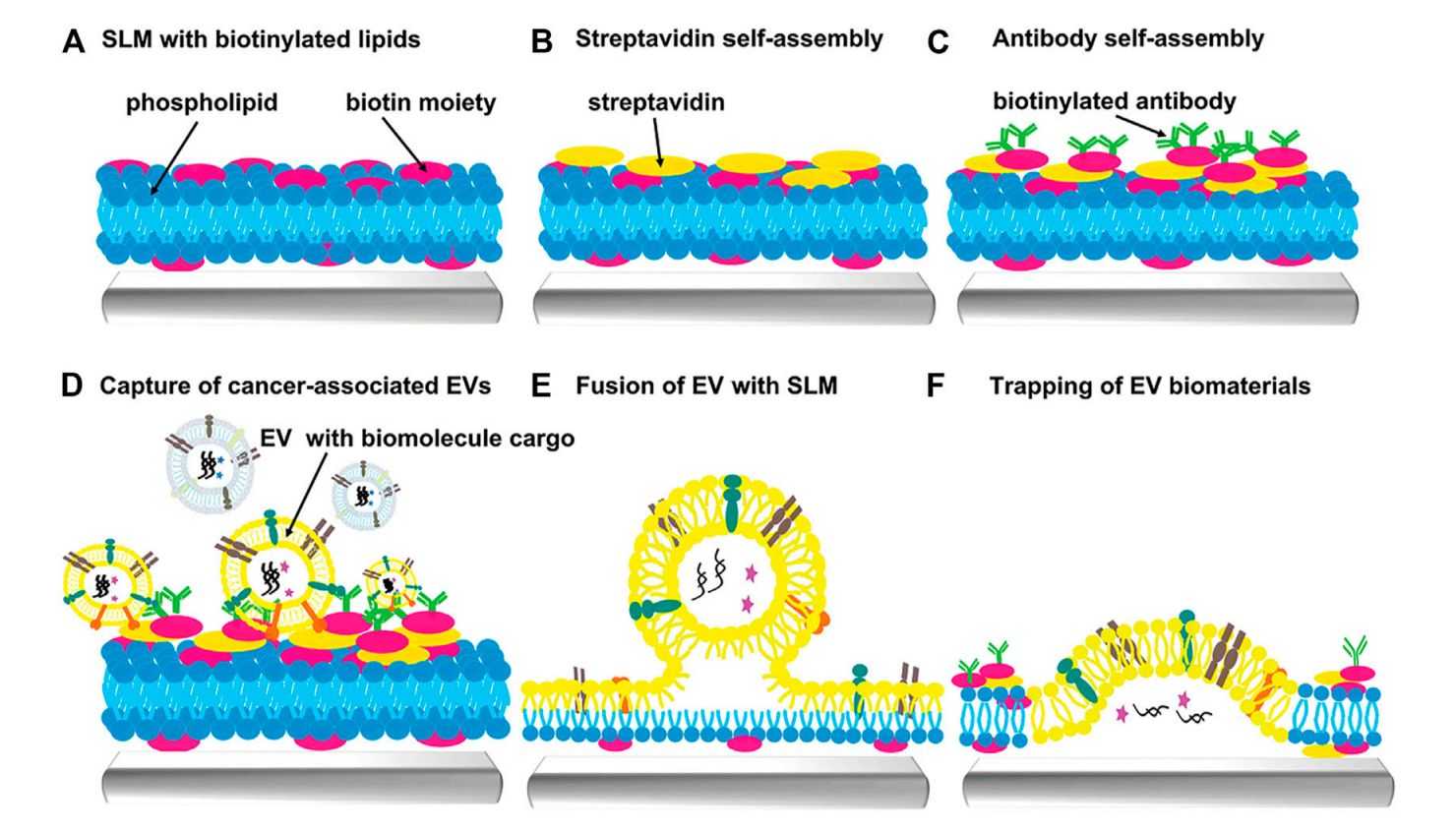
Since IA capture has the potential ability to distinguish between minimal conformational diversity. Researchers have successfully demonstrated that exosomes derived from different antigen-presenting cells exhibit specific combinations of biomarkers on their surfaces, and that immunocapture can recover the analyzed amount of vesicles on the surface of magnetic beads. These biomarkers are usually shared but are expressed at different levels in vesicles derived from different cell types.
In recent years, functionalized magnetic beads have been widely used for exosome isolation by combining microfluidic devices with immunological methods. Meanwhile, the integration of exosome isolation and detection can be realized by combining multiple methods. For example, label-free detection of specific proteins in exosomes based on surface plasmon resonance biochips provides effective information for cancer diagnosis and treatment.
 Figure 2. Scheme of extracellular vesicles capture by supporting lipid membranes (SLM). (Gao J, et al., 2023)
Why Choose IA Technology?
IA technology allows for the isolation of exosomes based on surface markers or antigens specifically of interest. This targeted enrichment facilitates the analysis of specific subpopulations of exosomes, allowing research into their unique characteristics, functional roles, and diagnostic or therapeutic potential. Exosomes isolated by IA technology are compatible with a variety of downstream analyses, including proteomics, lipidomics, and metabolomics, providing valuable insights into their roles and potential applications.
Creative Biostructure has been committed to the development and promotion of new exosome technologies. With our experienced team, we are more than happy to help clients with exosome-related research. If you are interested in new methods of exosome isolation, please feel free to contact us for more details and we will provide you with attentive service.
References
- Sonbhadra S, et al. Biogenesis, Isolation, and Detection of Exosomes and Their Potential in Therapeutics and Diagnostics. Biosensors. 2023. 13(8): 802.
- Gao J, et al. Recent developments in isolating methods for exosomes. Front Bioeng Biotechnol. 2023. 10: 1100892.
- Chen J, et al. Review on Strategies and Technologies for Exosome Isolation and Purification. Front Bioeng Biotechnol. 2022. 9: 811971.
- Popović, M., & de Marco, A. Canonical and selective approaches in exosome purification and their implications for diagnostic accuracy. Translational Cancer Research. 2017. 7(2): S209-S225.