Exosome Isolation by Microfluidics (MF)
Microfluidics (MF) is the science of researching the behavior, precise control, and manipulation of fluids and particles at the micron level, playing a critical role in the drug discovery process. Recently, scientists have also applied MF technology to isolate and detect exosomes. The MF platforms offer high-precision processing capabilities, the ability to handle microscale fluids, and integration with a variety of functional units. It can optimize the detection process on a single device and represents a powerful and versatile technology for exosome isolation and sensing, achieving high purity and high recovery in a short processing time.
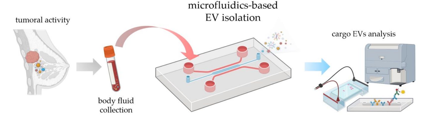 Figure 1. Possible workflow of tumor diagnosis using the microfluidic EV isolation strategy. (Meggiolaro A, et al., 2023)
What is the Principle of Microfluidic (MF) Technology?
- Microscale fluid manipulation - MF technology involves the precise manipulation and control of fluids at the microscale level. It utilizes microchannels, chambers, and various microstructures to manipulate fluid flow, mixing, and isolation.
- Size-based isolation - Exosomes typically range in size from 30-150nm. MF technology devices incorporate features such as microfilters, nanoporous membranes, or size-based capture structures to separate exosomes from larger particles such as cells, cellular debris, and protein aggregates.
- Continuous flow processing - MF-based exosome isolation is typically performed using continuous flow processing, in which the sample is introduced into the MF device and the fluid stream carries the exosomes through microstructures designed for isolation.
- Specific capture strategies - MF technology can be combined with specific capture strategies to enhance exosome isolation. For example, antibodies or aptamers targeting exosome surface labeling can be immobilized onto the MF surface or doped into the microstructure.
- Integration of downstream analysis - MF devices can be designed to integrate downstream analysis and processing steps directly into the MF platform. The transition from exosome isolation to subsequent characterization, such as proteomics, lipidomics, and metabolomics analyses, is achieved.
Advancement of Microfluidic-Based Exosome Isolation
- Microfluidic-based Exosome Isolation by Immunoaffinity
Exosomes can be pre-enriched from cell cultures or blood samples using immobilized antibodies on the MF surface. Researchers have designed an innovative exosome isolation device that allows for the isolation of CD63-specific exosomes from serum and simultaneous quantification and characterization. In addition to modifying chip surfaces or beads with CD63 to capture specific exosomes, alternative methods of targeting particular exosomal lipids have recently been researched. Examples include the membrane-bound protein V immobilized MF device integrated with an alternating 3D corrugated structure, which achieves up to 90% separation efficiencies for isolating cancer-derived exosomes.
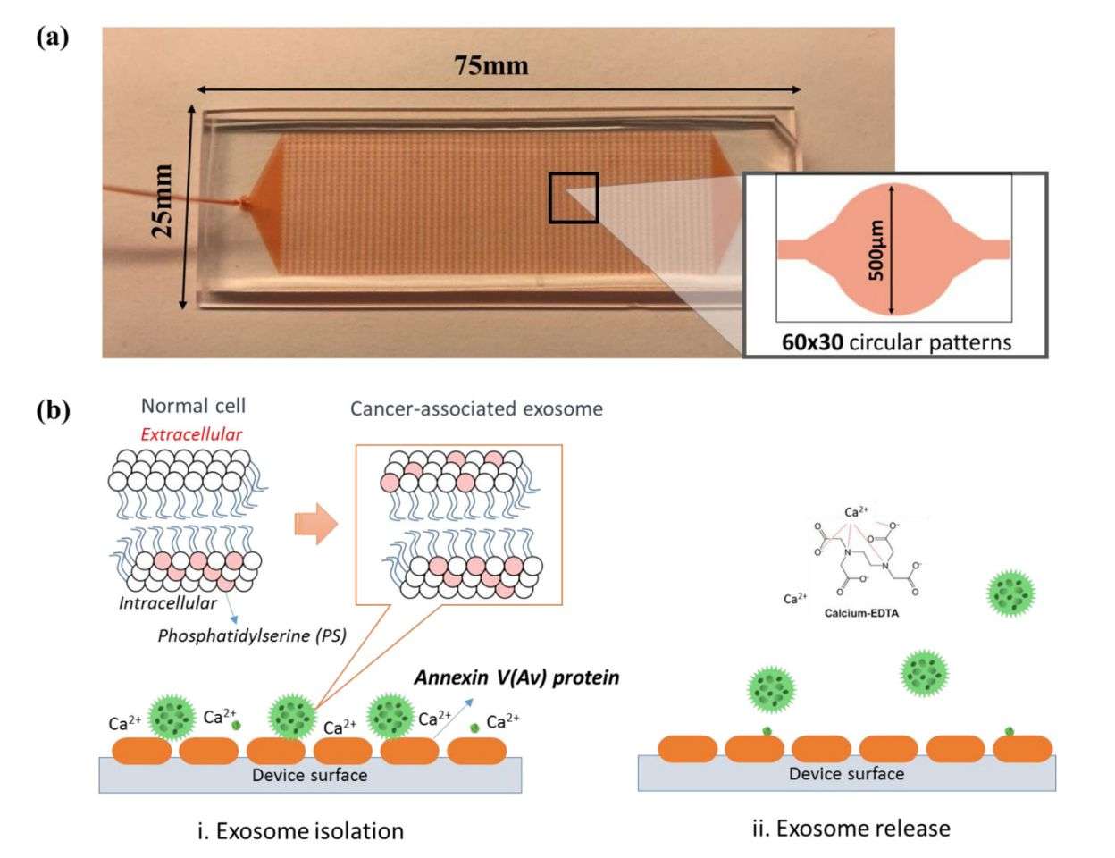 Figure 2. 3D ripple-like structure chip for the immunocapture and release of exosomes. (Kang YT, et al., 2019)
- Microfluidic Device for Label-Free Separation of Exosomes
MF-based filtration for the separation of exosomes is similar to traditional ultrafiltration methods involving the use of nanomembranes or nanowires. Researchers applied the novel structure of porous silicon nanowires to separate exosomes between 40-100nm. Using a similar strategy, a 3D PDMS anchored with ZnO nanowires was designed to enable exosome capture in the device. A powerful tool for improved early diagnosis of urine-related diseases. In addition, the scientists used hydrodynamic properties, acoustic waves, and electrical properties to separate exosomes.
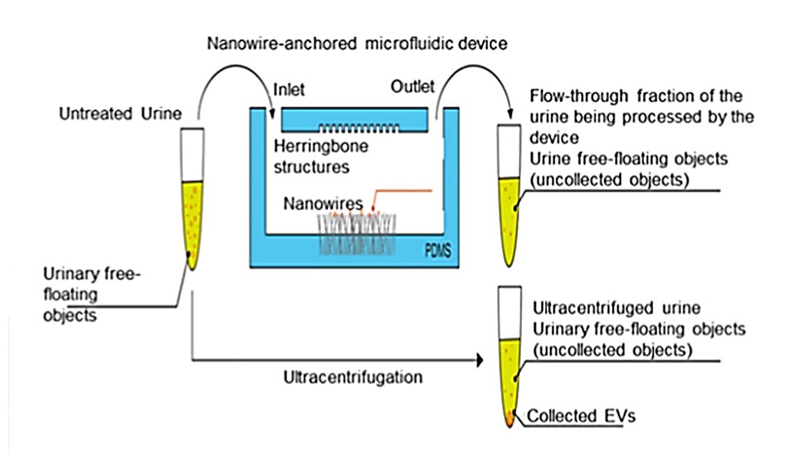 Figure 3. Schematic for the exosome isolation from urine using a microfluidic device anchored with ZnO/Al2O3 core-shell nanowire. (Yasui T, et al., 2017)
Benefits of MF Technology
- Consumption of small volumes of reagents
- Improvement of mass and heat transfer
- Laminar flow
- Parallelization
- Portability
- High throughput and multiple analysis
- Low power consumption
- Faster analysis and quick response
- Rapid screening of nanoparticles
- Continuous flow operations
Creative Biostructure is an expert in the efficient isolation of exosomes, and we provide reliable exosome isolation services and high-quality products to our clients. Our products include exosome kits and high-purity exosomes for biomarker discovery and drug delivery research.
| Cat No. | Product Name | Source |
| Exo-UEK01 | Urine Exosome Kit | / |
| Exo-HDBF-01 | HQExo™ Exosome-SDH-Alzheimer's plasma | Exosome derived from Single Donor Human Alzheimer's plasma |
| Exo-HDBF-02 | HQExo™ Exosome-SDH-Asthma plasma | Exosome derived from Single Donor Human Asthma plasma |
| Exo-BF01 | HQExo™ Exosome-CSF | Exosome derived from human pooled CSF (healthy donors) |
| Exo-BF12 | HQExo™ Exosome-Human biofluids ascites fluid | Exosome derived from Human Ascites Fluid (healthy donors) |
| Exo-CM01 | HQExo™ Exosome-B16F10 | Exosome derived from murine melanoma cell line (B16F10 cell line) |
| Exo-CH15 | HQExo™ Exosome-BLCL21 | Exosome derived from EBV transformed lymphoblastoid B cells (BLCL21 cell line) |
| Explore All Exosome Products | ||
In addition, we offer a range of downstream research including exosome characterization, labeling, tracking, and functional analysis. If you are interested in our services, contact us today to explore the possibilities that exosome isolation services can bring to your research.
References
- Meggiolaro A, et al. Microfluidic Strategies for Extracellular Vesicle Isolation: Towards Clinical Applications. Biosensors. 2023. 13(1): 50.
- Kang YT, et al. Isolation and Profiling of Circulating Tumor-Associated Exosomes Using Extracellular Vesicular Lipid-Protein Binding Affinity Based Microfluidic Device. Small. 2019. 15(47): e1903600.
- Yasui T, et al. Unveiling massive numbers of cancer-related urinary-microRNA candidates via nanowires. Sci Adv. 2017. 3(12): e1701133.
- Wang J, et al. Towards Microfluidic-Based Exosome Isolation and Detection for Tumor Therapy. Nano Today. 2021. 37: 101066.
- Verma A, Bhattacharyya S. Microfluidics - The State-of-the-Art Technology for Pharmaceutical Application. Adv Pharm Bull. 2022. 12(4): 700-711.

