Dynamic Light Scattering (DLS) for Exosome Characterization
Exosomes are a class of extracellular vesicles containing mRNAs, miRNAs, and proteins that can control the properties of other cells or the surrounding environment. Increasing evidence of carcinogenicity of tumor-derived exosomes has attracted widespread attention. Over the decades, there has been a proliferation of techniques to accurately characterize exosomes, including dynamic light scattering (DLS).
Dynamic light scattering (DLS, also known as photon correlation spectroscopy or quasi-elastic light scattering) is the most commonly used measurement technique for particle size analysis in the nanometer range. In recent years, DLS has allowed scientists to monitor the release of smaller exosomes from healthy and dying cells.
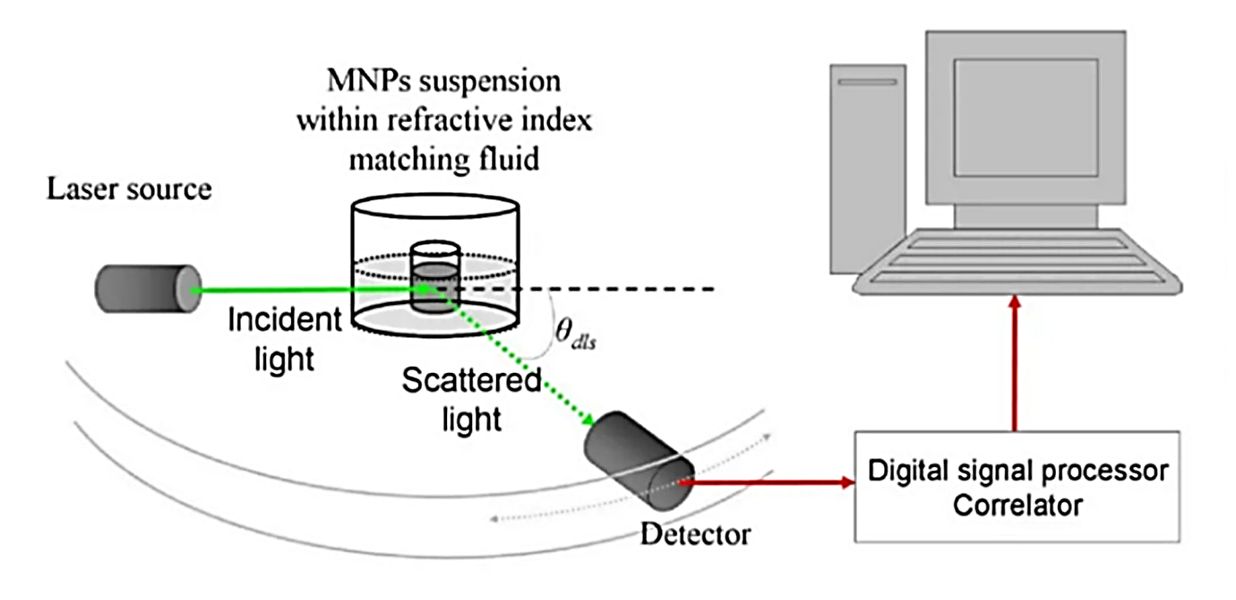
 Figure 1. A schematic of the dynamic light scattering (DLS) setup. (Kouhpanji MRZ, Stadler BJH, 2020)
What is the Principle of DLS?
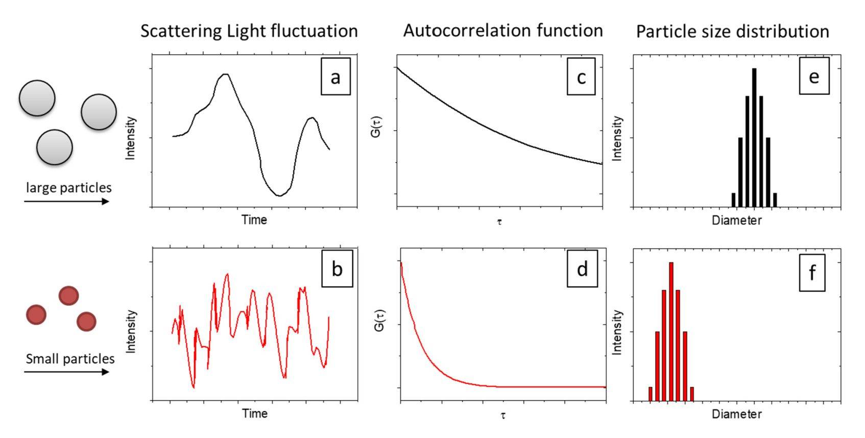
DLS is based on the Brownian motion of dispersed particles. When particles are dispersed in a liquid, they move randomly in all directions. The principle of Brownian motion is that the particles are constantly colliding with solvent molecules. These collisions result in a certain amount of energy transfer, which causes the particles to move. The energy transfer is more or less constant and has a greater effect on smaller particles. As a result, smaller particles move at a higher speed than larger particles. The hydrodynamic diameter can be determined by measuring the velocity of the particles if all other parameters affecting particle motion are known.
 Figure 2. Principle diagram of dynamic light scattering (DLS). (Jia Z, et al., 2023)
In the context of DLS, time fluctuations are usually analyzed by intensity or photon autocorrelation function (ACF). A monochromatic beam of light (e.g., a laser) is directed into a test solution containing spherical particles moving in Brownian motion, and when the light strikes the moving particles it induces a Doppler shift, which changes the wavelength of the original light. This change is related to the size of the particle. By measuring the diffusion coefficient of the particles in the test medium by ACF, the size distribution of the spheres can be calculated and the motion of the particles in the test medium can be described in detail. Meanwhile, DLS can also be used to probe the behavior of complex fluids, such as concentrated polymer solutions.
DLS Analyzing Steps

1. Sample Preparation - Clean the sample bottle or test cell to ensure that it is free of contaminants, dust, or scratches. Prepare sample solutions with attention to the degree of dilution, choice of solvent, and homogeneity of the sample. Clients provide exosomes or samples (we provide exosome isolation service).
2. Equipment Calibration - Perform system calibration according to the requirements of the instrument, including temperature settings, software setup, and initial calibration using standard particles.
3. Sample Loading - Load the sample solution into the test cell or sample vial, making sure there are no air bubbles.
4. Parameter Setting - Set the appropriate parameters on the DLS instrument, such as measurement time, temperature, and laser intensity. These parameters need to be adjusted according to the characteristics of the sample.
5. Perform Measurement - Initiate the measurement. The instrument records the time variation of the scattered light intensity and analyzes this data to determine the size of the particles.
6. Data Analysis - The data is analyzed using the instrument software. The DLS typically provides information on the particle size distribution, including the average particle size and the width of the distribution. For complex samples, it may be necessary to make multiple measurements or use different analytical models to obtain accurate results.
What are the Precautions for Using DSL?
- Data Quality
The data quality of DLS measurements has factors in the instrument hardware, such as the quality of the laser, detector, and correlator. In addition, the following factors significantly affect the quality of the measurement and need to be taken into account by the tester.
a) Scattering angle
The chirp rate is related to the wave vector. The intensity of scattering varies with the scattering angle to different degrees for different particle sizes. Therefore there is a better detection angle for each particle size. To ensure a high-quality test and analysis, the test needs to be performed at multiple angles.
b) Multiple scattering
DLS is only applicable to single scattered light and data interpretation becomes very difficult for systems with non-negligible multiple scattering contributions. Especially for larger particles with high scattering contrast, the upper concentration limit is even lower. As a result, many systems are excluded from the research of conventional dynamic light scattering. However, multiple scattering in DLS can be suppressed by a mutual correlation approach. The design concept is to isolate single-scattered light and suppress contributions from undesired multiple scattering in DLS experiments.
- Data Analysis
All particles in a solution can't have the same hydrodynamic radius Rh, which usually has a distribution. Its size and distribution width vary from sample to sample. The width of the distribution leads to a distribution of diffusion coefficients and consequently a distribution of the chirp rate Γ of g1(τ). Thus g1(τ) does not simply decay exponentially.
Applications
- Liposome and exosome characterization
- Protein stability analysis
- Formulation testing
- Nanoparticle characterization
- Polymer and macromolecule analysis
- Aggregation assays
Why Choose DLS?
- Accurate, reliable, and reproducible particle size analysis.
- Simple sample preparation, even natural samples can be analyzed directly without sample preparation.
- Low volume requirements.
- Simple setup and fully automated measurement.
- Measures sizes <1nm.
- Measure molecules with molecular weights <1000Da.
Creative Biostructure specializes in exosome characterization services using cutting-edge dynamic light scattering (DLS) technology. With years of experience in nanoparticle analysis, we can provide precise and accurate results to support scientific research and product development. Whether you are engaged in drug delivery research, biomarker research, or developing nanomedicines, our services provide the comprehensive analysis to move your project forward. Please feel free to contact us for more details.
References
- Kouhpanji MRZ, Stadler BJH. A Guideline for Effectively Synthesizing and Characterizing Magnetic Nanoparticles for Advancing Nanobiotechnology: A Review. Sensors (Basel). 2020. 20(9): 2554.
- Jia Z, et al. Dynamic Light Scattering: A Powerful Tool for In Situ Nanoparticle Sizing. Colloids and Interfaces. 2023. 7(1): 15.
- Falke S, Betzel C. Dynamic Light Scattering (DLS): Principles, Perspectives, Applications to Biological Samples. Radiation in Bioanalysis. 2019. 8: 173-193.