Cryo-EM for Nanomaterials
Cryogenic electron microscopy (cryo-EM) technology is generally used to observe temperature-sensitive samples, such as proteins and nanomaterials. They are flash-frozen to reduce the damage of electron beam and avoid particle deformation. In this way, a more real sample morphology can be obtained. Cryo-EM has many advantages, including high acceleration voltage, good electronic and optical performance, automatic process, and so on. In addition to life sciences, cryo-EM has also provided opportunities in materials science by enabling higher resolution characterization of temperature-sensitive materials.
Nanomaterials, usually means that materials of which a single unit is sized between 1 to 100 nm. Nanomaterials including the biological material (such as bacteria and viruses), some inorganic and organic compounds. Aqueous nanomaterials can form stable structure in the solvent, but after drying in the air, it has been great changes in the structure form. Therefore, in the use of conventional negative staining technique cannot get the real structure. For the unstable nanomaterials in a vacuum or in the air, due to there is no need a lot of particles and the complicated calculation, single particle Cryo-EM technology is relatively simple and can provide a structure in a native state, which makes Cryo-EM becoming an essential technology for studying the structure of nanomaterials.
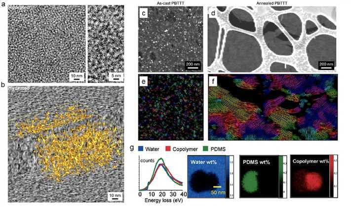 Figure 1. Cryo-EM for soft polymer materials. (Li Y
et al
., 2020).
Figure 1. Cryo-EM for soft polymer materials. (Li Y
et al
., 2020).
Cryo-EM also plays a role in observing the in-situ structures of complex or organelles in living cells. It is suitable to study the biomolecules and their composites that do not have uniformity at nanoscale. At the same time, compared with the negative-staining EM method, cryo-EM can avoid the defects such as dye illusion and vacuum deformation, and can reflect the original structure characteristics of the nanomaterial samples.
Our cryo-EM team has rich experience in nanomaterial, subcellular organelles, filaments, biological tissues, and other EM samples. We are happy to customize the cryo-EM process of nanomaterial based on your requirements.
If you are interested in our high-resolution nanomaterial characterization service, please feel free to contact us . We are looking forward to cooperating with you.
Ordering Process

Reference
- Li Y, et al . Opportunities for cryogenic electron microscopy in materials science and nanoscience. ACS Nano . 2020. 14(8): 9263-9276.

