Exosome
Exosomes are nano-sized small extracellular vesicles secreted by cells, which can carry nucleic acids, proteins, lipids, and other bioactive substances. They have been playing an important role in the physiological and pathological processes.
Establishing the method to capture, identify, and quantify exosomes is the premise of clinical testing. At present, the most commonly used methods for exosome separation and enrichment are ultra-centrifugation, polymerization precipitation, immunoaffinity, ultrafiltration, and size-exclusion chromatography. The isolated exosomes can be detected by a variety of electron microscopes, such as scanning electron microscopy (SEM), transmission electron microscopy (TEM), atomic force microscopy (AFM), and cryo-electron microscopy (cryo-EM). Our company can perform exosome analysis with all of these different methods, especially cryo-EM.
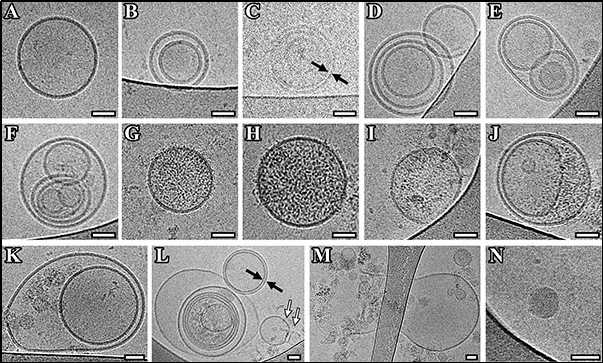
 Figure 1. Cryo-EM images of extracellular vesicles (EVs) isolated from pooled CSF of Parkinson's disease patients (Emelyanov A, et al. 2020)
Compared with synthetic vectors such as liposomes and nanoparticles, exosomes have extensive and unique advantages in the field of disease diagnosis and treatment due to their internality and heterogeneity. Our cryo-EM platform adopts the latest ultra-high resolution cryo-EM mode, which enables fast sample processing, rapid embedding, and clear morphology imaging of exosome samples.
If you are interested in our exosome particle morphology analysis service, please feel free to contact us. We are looking forward to cooperating with you.
Ordering Process
Reference
- Emelyanov A, et al. Cryo-electron microscopy of extracellular vesicles from cerebrospinal fluid. PLoS One. 2020. 15(1): e0227949.

