4D UEM Service
The discovery of the electron over a century ago and the realization of its dual character have given birth to one of the two most powerful imaging instruments: the Electron Microscope (EM). The EM’s ability to resolve three-dimensional (3D) structures on the atomic scale is continuing to affect different fields, including materials science and biology. In addition to the progress made in increasing the spatial resolution, the Transmission Electron Microscope (TEM) has become an all-in-one characterization tool. In conventional TEMs, electrons are produced by heating a source or by applying a strong extraction field. Both methods result in the stochastic emission of electrons, with no control over temporal spacing or relative arrival time at the specimen. Today, ultrafast electron microscopy (4D UEM) enables a resolution that is 10 orders of magnitude better than that of conventional microscopes.
Based on our unparalleled in-house EM platform, Creative Biostructure provides high-speed 4D UEM diffraction analysis, which is a benefit of a high-speed camera. The fast frame rate camera was applied to drive the UEM probe on beam sensitive ZnO NWs, grown on GaN substrates. High-speed 4D UEM data acquisition significantly reduced the specimen damage previously encountered during data collection (saving time and money). The data acquired with the camera provides high-resolution information capable of direct observation of single systems over large length-scales. Through intelligent modeling of this data, it also enables a visualization of nanoscale effects in an overall microsystem. Such results produce insights into the mechanisms of nano epitaxial growth and foundational information to incorporating these systems into future technologies.
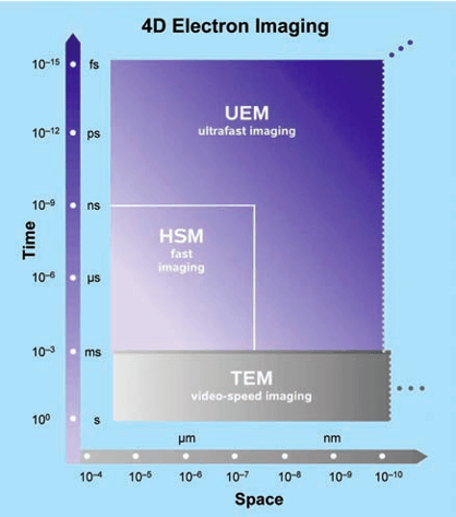
 Figure 1. 4D electron imaging. The resolution boundaries of ultrafast imaging are compared with those achieved in conventional TEM, limited by the speed of video camera, and, in high-speed microscopy (HSM), defined by the rectangle shown. The spatiotemporal scales of UEM achieved to date are outlined with possible future extensions.
In UEM space-time domains (Figure 1), images are obtained stroboscopically with single-electron coherent packets. Under such a condition, electron repulsion is absent, permitting real-space imaging, Fourier-space diffraction, and energy-space electron spectroscopy with high spatiotemporal resolutions. The time resolution becomes limited only by the laser pulse width and energy width of the packets, the camera rate of recording becomes irrelevant for the temporal resolution, and the delay between pulses can be controlled to allow for the cooling of the specimen and/or repetitiveness of the specimen’s exposure.
The applications to display phenomena of different length and time scales:
- Atomic motions in structural dynamics
- Phase transitions
- Membrane mechanical drumming
The facility in Creative Biostructure is home to world class equipment including negative staining EM, SEM and cryo-TEM that enable our customers to complete in-depth investigations of material properties, including crystallinity, defects, and microstructure. We strive to provide you expertise necessary for success in your research and development projects.
We encourage you to contact us to discuss how we can be of assistance to you.
Ordering Process
References
- David J. Flannigan and Ahmed H. Zewail. 4D Electron Microscopy: Principles and Applications. ACCOUNTS OF CHEMICAL RESEARCH. 1828–1839, 2012, Vol. 45, No. 10.
- Ahmed H. Zewail. Four-Dimensional Electron Microscopy. Science 328, 187 (2010).
