Exosomes Isolated from Cancer Cell Lines
Exosomes are a vital part of intercellular communication and have garnered significant attention in cancer research for their role in cancer progression, diagnostics, and therapy. At Creative Biostructure, we offer high-quality exosome standards derived from various cancer cell lines, suitable for a range of research applications including WB, ELISA, FACS, and more. These exosomes serve as control standards and can be used for quantitative calibration of exosomes in biological samples. Our services cater to both academic and industrial research needs, with options for customized exosome isolation to meet specific requirements.
Product List
Background
Exosomes are small, membrane-bound extracellular vesicles (EVs) ranging from 40 to 160 nm in size. They are secreted by almost all cell types, including cancer cells, and play a critical role in the regulation of many physiological and pathological processes. In cancer research, exosomes have emerged as key mediators of tumor metastasis, immune response modulation, and drug resistance. Exosomes carry a diverse array of bioactive molecules such as proteins, nucleic acids, lipids, and metabolites, which reflect the molecular state of their cell of origin. This cargo can influence distant cells and organs, making exosomes ideal for liquid biopsies and non-invasive cancer diagnostics.
The exosomes isolated from cancer cell lines offer unique insights into the tumor microenvironment and the molecular signatures that drive cancer progression. These exosomes are particularly advantageous for cancer research due to their ability to carry oncogenic proteins, RNAs, and lipids that can reprogram recipient cells. The robust secretion of exosomes by cancer cells also enhances their utility for research, providing an abundant and reliable source of material for experimental studies.
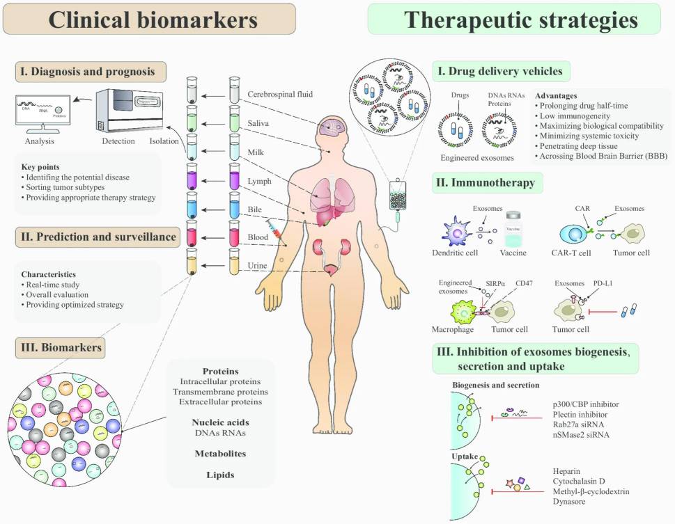 Figure 1. Clinical applications of exosomes in cancer. (Zhu L, et al., 2020)
Applications of Exosomes in Cancer Research
Cancer Diagnostics
Exosomes are abundant in bodily fluids and carry specific biomarkers reflective of the cancerous state of their originating cells. These molecules, including tumor-specific proteins, DNA, and RNA, can serve as biomarkers for early cancer detection. The isolation of exosomes from cancer cell lines allows researchers to study these biomarkers in vitro. This provides a powerful tool for understanding tumor progression and developing diagnostic assays for non-invasive liquid biopsies.
Therapeutic Target Discovery
Exosome-mediated intercellular communication plays a significant role in tumor progression, making exosomes an important target for cancer therapies. We provide exosome isolation services that enable researchers to study the specific cargo of exosomes derived from cancer cells. This cargo includes key proteins, microRNAs, and other molecules involved in processes such as immune evasion, metastasis, and drug resistance. Understanding these molecular mechanisms is critical for identifying therapeutic targets aimed at disrupting the exosome-mediated pathways that contribute to cancer.
Drug Delivery Research
The natural ability of exosomes to transfer bioactive molecules to specific target cells makes them promising vehicles for drug delivery. Researchers are exploring the use of cancer-cell-derived exosomes to deliver anti-cancer drugs or therapeutic RNAs directly to tumor cells, thus minimizing off-target effects and improving treatment efficacy. Creative Biostructure offers customized exosome products to facilitate the exploration of exosome-based drug delivery systems for cancer treatment, including the development of exosome engineering techniques that enhance targeting specificity and drug loading capacity.
Case Studies
Case Study 1: Exosome-Derived KRAS Mutations as Early Biomarkers in Pancreatic Cancer Detection
This study highlights the utility of exosome-derived DNA (exoDNA) in detecting KRAS mutations in patients with pancreatic ductal adenocarcinoma (PDAC). It was found that exoDNA provides a higher detection rate of KRAS mutations in early-stage PDAC patients compared to circulating cell-free DNA (cfDNA), with 66.7% of localized PDAC patients showing KRAS mutations in exoDNA, compared to only 45.5% in cfDNA. These findings underscore the potential of exoDNA as a valuable tool in early cancer detection and prognostic stratification, offering a complementary source to cfDNA in liquid biopsies. However, KRAS mutations were also detected in a small percentage of healthy individuals, suggesting the need for caution in interpreting these results for cancer screening.
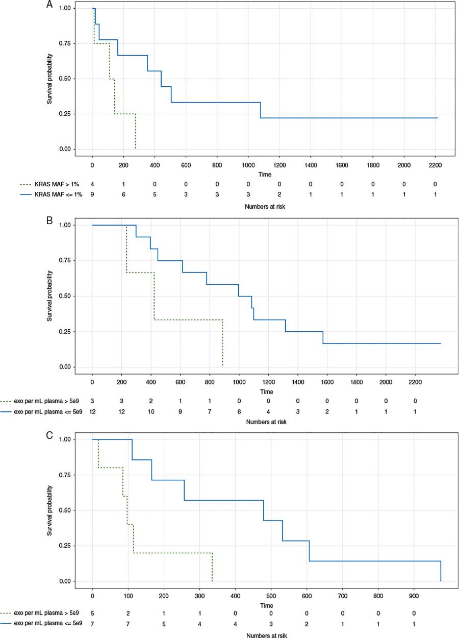 Figure 2. Impact of KRAS mutant allele frequency and exosome concentration on disease-free and overall survival in pancreatic cancer patients. Kaplan-Meier survival curves showing the influence of (A) KRAS mutant allele frequency (MAF) and (B, C) exosome concentration on patient survival in pancreatic ductal adenocarcinoma (PDAC). Panel (A) compares disease-free survival between patients with KRAS MAF greater than 1% and less than or equal to 1%. Panels (B) and (C) illustrate overall survival in patients with localized and metastatic PDAC based on exosome concentration, with higher exosome levels correlating with shorter survival times. (Allenson K, et al., 2017)
Case Study 2: Effects of Different Isolation Methods on the Purity and Yield of Melanoma Exosomes
This study compares different exosome isolation methods, such as ultracentrifugation, size-exclusion chromatography (SEC), and a combined ultrafiltration and SEC method, focusing on melanoma-derived exosomes. The findings show that the combined ultrafiltration and SEC method significantly improves exosome purity and yield, providing up to 58-fold more exosomes and reducing co-isolated soluble factors by 836-fold compared to ultracentrifugation. This method also isolated 2.5 times more PD-L1-expressing exosomes, which are critical in studying melanoma's immune evasion mechanisms.
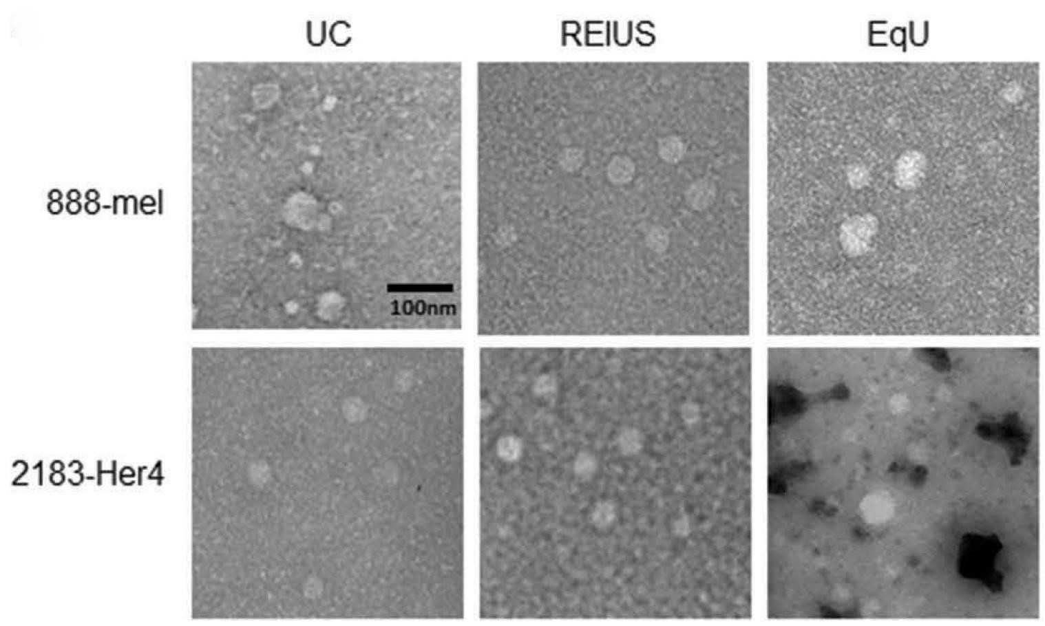 Figure 3. Transmission electron microscopy of isolated melanoma exosomes. Negative staining transmission electron microscopy images of exosomes isolated from melanoma cell lines (888-mel and 2183-Her4) using various methods. Exosomes display a circular morphology with intact membranes, confirming successful isolation. (Shu S L, et al., 2020)
Product Advantages
- High Purity: Our advanced isolation techniques ensure the production of high-purity exosomes with minimal contamination from non-exosomal proteins, other extracellular vesicles, or cellular debris. This level of purity is critical for experiments that require precise molecular analyses, such as proteomics, RNA sequencing, or lipidomics.
- Stable Bioactivity: Exosomes retain their biological activity after isolation, maintaining the integrity of their protein, lipid, and RNA cargo. This ensures that the exosomes used in research reflect the true physiological functions of cancer-derived exosomes, allowing for accurate investigation of their roles in cancer biology.
- Precise Exosome Characterization: Each exosome preparation is rigorously characterized by size, morphology, and molecular content. We utilize state-of-the-art techniques, including nanoparticle tracking analysis (NTA), transmission electron microscopy (TEM), and flow cytometry, to ensure that exosomes meet the highest standards of quality. These characterizations provide researchers with the detailed information needed for reproducible and high-impact studies.
- Customized Solutions: We work closely with our clients to design personalized exosome isolation protocols that meet their specific research goals. Whether you need exosomes from a specific cancer cell line or require particular modifications for your studies, we provide customized solutions to ensure success.
Resources
Frequently Asked Questions
-
How do you ensure the purity of your exosome preparations?
We use advanced isolation techniques, such as ultracentrifugation, size-exclusion chromatography, and immunoaffinity capture, to produce exosomes with the highest possible purity.
-
Can I request exosomes from a specific cancer cell line?
Yes, we offer customized exosome isolation services. We can isolate exosomes from various cancer cell lines based on your research requirements.
-
Are your exosome products suitable for drug delivery research?
Yes, our exosome products are ideal for studying drug delivery mechanisms. We can also offer engineered exosomes to suit specific research needs.
Contact us today to learn more about how our exosome products can accelerate your cancer research initiatives!
References
- Allenson K, Castillo J, San Lucas F A, et al. High prevalence of mutantKRAS in circulating exosome-derived DNA from early-stage pancreatic cancer patients. Annals of Oncology. 2017. 28(4): 741-747.
- Shu S L, Yang Y, Allen C L, et al. Purity and yield of melanoma exosomes are dependent on isolation method. Journal of Extracellular Vesicles. 2020. 9(1): 1692401.
- Zhu L, Sun H T, Wang S, et al. Isolation and characterization of exosomes for cancer research. Journal of Hematology & Oncology. 2020. 13: 1-24.
