Exosome In Vivo Functional Assay
Exosomes (30-160 nm in diameter), a major type of secreted extracellular vesicles (EVs), are derived from intraluminal vesicles (ILVs) in the early endosomal compartment. Exosomes are small lipid bilayer vesicles produced by most cells, which contain abundant nucleic acids, proteins and lipids. Exosomes have been widely used in the research of tumor therapy drugs because of their good biocompatibility, circulation ability in vivo and material delivery capacity.
Exosomes are biological nanoparticles, which have immense prospects of clinical application as a diagnostic marker, and can be used in monitoring the disease progression and as the potential therapeutic carrier. Creative Biostructure can provide a variety of in vivo functional tests for exosome functional assays, such as exosome reporter mice, subcutaneous tumor formation, in vivo imaging, immunofluorescence, etc.
- Exosome Reporter Mice
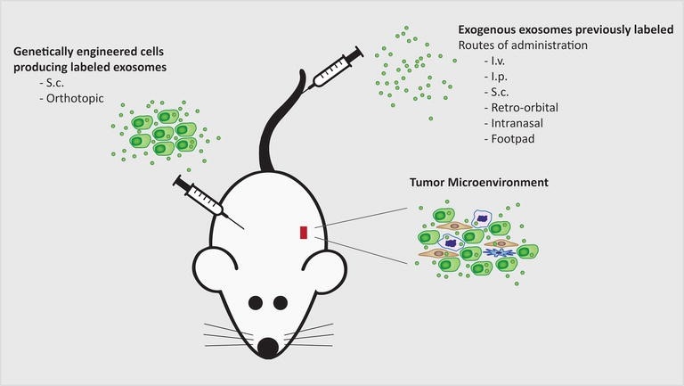
Genetically engineered mouse models (GEMM) could be a promising approach to trace tumor-derived exosomes in a living organism. Our common exosome markers are several tetraspanin family proteins (CD81, CD9, and CD63) and TSG101, Alix, etc. In addition, there are many methods to prepare exosome reporter mice: fluorescent labeling and bioluminescence reporters. Among them, exosome labeled with fluorescent is the most widely used method to trace their fate in vivo.
 Figure 1. Different methods available to prepare exosome reporter mice. (Bárbara Adem, et al. 2016)
Exosome Reporter Mice Preparation Process:

- Subcutaneous Tumor Formation Assay
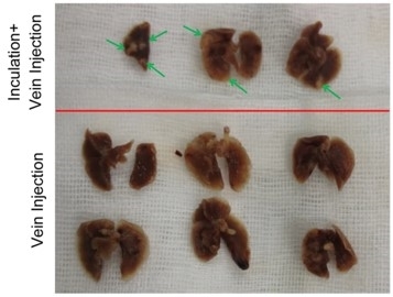
A subcutaneous injection is a method of administering medication. It had been reported that subcutaneous injection of exosomes reduces symptom severity and mortality induced by Echinostoma Caproni infection in BALB/c mice.
 Figure 2. Primary tumor-derived exosomes were intravenously injected into mice. (Fu, Q., et al. 2018)
- In vivo Imaging
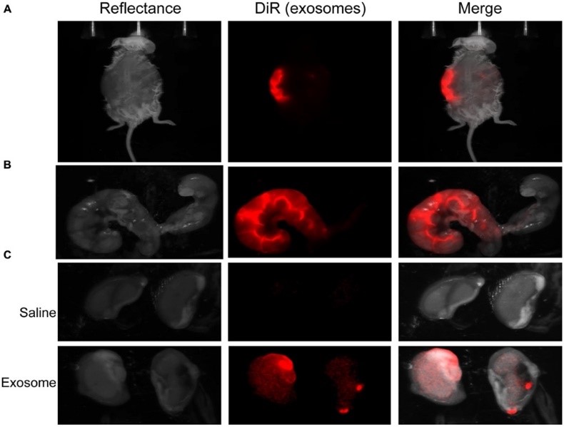
To determine the trafficking of exosomes injected into the body, we provide animals imaging after injection. Fluorescently labeled exosomes were injected into mice and imaged after 24 h. exosome-injected mouse images showed fluorescence.
 Figure 3. In vivo imaging of pregnant mouse 24 h post injection. (Sheller-Miller, S., et al. 2016)
- Immunofluorescence
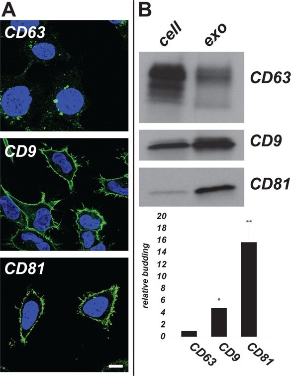
Using immunofluorescence (IF) microscopy, we provide the variations in exosome composition. We provide multiple exosome marker antibodies, such as CD9 and CD81, to meet your needs for WB, IHC, IF, etc.
 Figure 4. Plasma membrane-localized exosome cargoes CD9 and CD81 display higher relative budding than endosome-localized CD63. (Francis K. Fordjour, et al. 2019)
If you are interested in our exosome in vivo functional assay, please feel free to contact us. We are looking forward to cooperating with you.
Ordering Process
References
- Bárbara Adem and Sónia A. MeloBusatto. Animal models in exosomes research: what the future holds. Novel Implications of Exosomes in Diagnosis and Treatment of Cancer and Infectious Diseases. 2016, chapter 3.
- Fu, Q., et al. Primary tumor-derived exosomes facilitate metastasis by regulating adhesion of circulating tumor cells via SMAD3 in liver cancer. Oncogene. 2018, 37: 6105-6118.
- Sheller-Miller, S. et al. Feto-maternal trafficking of exosomes in murine pregnancy models. Frontiers in Pharmacology. 2016, 7: 432.
- Francis K. Fordjour, et al. A shared pathway of exosome biogenesis operates at plasma and endosome membranes. BioRxiv. 2019.

