X-ray Diffraction Data Collection for Coronavirus Research
The quality of X-ray diffraction data of the protein crystal can determine the success rate of the subsequent steps of structure determination and the reliability of the reconstructed three-dimensional (3D) structural model. After successfully obtaining high-quality crystals for X-ray diffraction, Creative Biostructure supports the collection of X-ray diffraction data to facilitate protein structural analysis and drug candidate screening involved in the research of coronavirus infection.
Introduction to Coronavirus and SARS-CoV-2 Infection

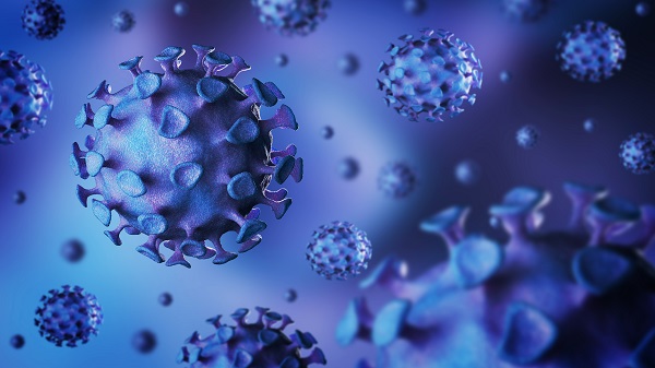
Novel coronavirus (also known as SARS-CoV-2) is an enveloped β-coronavirus with a positive-sense single-stranded RNA genome. Similar to the other two coronaviruses that cause SARS (severe acute respiratory syndrome) and MERS (Middle East respiratory syndrome), its genome encodes non-structural proteins, structural proteins, and helper proteins. Non-structural proteins include 3-chymotrypsin-like protease (3CLpro), papain-like protease (PLpro), helicase, and RNA-dependent RNA polymerase (RdRp). Structural proteins include the spike glycoprotein (S protein). Currently, the global academia and biotech/pharmaceutical industries are striving to develop coronavirus-specific antiviral drugs and vaccines that can be used to treat or prevent SARS-CoV-2 infection.
X-ray Diffraction Data Collection for Coronavirus Research
Creative Biostructure utilizes the powerful in-house Rigaku X-ray equipment (for testing and optimization) and synchrotron radiation (for formal data collection) to determine high-resolution structures. Our crystallographers can improve diffraction quality by optimizing cryoprotectants, annealing agents, and dehydrants.
Different data collection methods can affect the data quality, which is closely related to the reliability of the final 3D-structure determination. We can collect multiple data sets according to different phase determination methods, which include molecular replacement (MR), multiple isomorphous replacement (MIR), and single-/multi-wavelength anomalous dispersion (SAD/MAD). If there is an experimental structure of the homologous protein, we can collect a set of diffraction data for MR; In the absence of homologous structures, we support the MIR method by collecting data sets of the crystal after soaking in a heavy atom solution or the SAD/MAD method of selenomethionine (SeMet) derivatives to determine the phase. Depending on the method utilized, more than one set of data can be collected.
Why Do You Choose Us?
- Advanced contract service provider in the field of crystallography, with experience in the structural analysis of biomacromolecules related to virus research
- Services are unbundled, the crystal used for X-ray diffraction data collection can be provided by our customer or obtained from our crystallization service
- Providing detailed X-ray diffraction data report
- The project information and diffraction data are strictly confidential
- Our customer service representatives are available 24 hours a day from Monday to Sunday
Contact us to discuss your project!


