Scanning Electron Microscopy for Coronavirus-like Particle
Scanning electron microscopy (SEM) is characterized by simple sample preparation, wide adjustable magnification range, high image resolution, and large depth of field. SEM technology can scan the surface morphology of the sample by emitting a bright focused electron beam. The electron beam interacts with the appearance of the sample surface to generate a variety of signals that can be detected. These signals include the surface morphology and composition of the sample. Creative Biostructure provides SEM services for the characterization and research of coronavirus and coronavirus-like particles.
Application of SEM in Coronavirus Research
Novel coronavirus (SARS-CoV-2, formerly known as 2019-nCoV) causes COVID-19 pneumonia, which has become a global public health emergency since it was first discovered in Wuhan, China, in December 2019. Using scanning and transmission electron microscopy, scientists obtained a series of ultrastructure images of the novel coronavirus. This method has become a reliable tool for classifying viruses and clarifying their structure and function according to their ultrastructure. The electron microscopic image of the novel coronavirus does not look quite different from the original severe acute respiratory syndrome coronavirus (SARS-CoV, appeared in 2002) and the Middle East respiratory syndrome coronavirus (MERS-CoV, appeared in 2012). The crown-like spike structure on the surface of the virus explains the name of the Coronaviridae family, and most coronaviruses have a crown-like appearance.
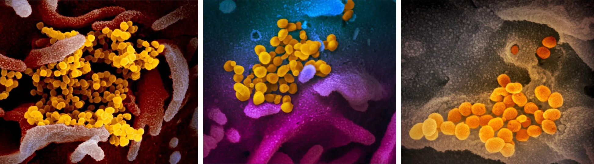 Figure 1. Scanning electron microscope images show SARS-CoV-2 (yellow). (photo source: NIAID-RML)
Our SEM Services for Coronavirus-like Particle
With our state-of-the-art high-resolution field emission scanning electron microscope, we can satisfy your pursuit of high-quality experiments. Our services are not for diagnostic testing, only for research purposes, and we do not support the observation of natural coronaviruses capable of causing large-scale human infections. The customer only needs to provide us with tissues or cell cultures, and we will eventually deliver high-resolution scanning imaging results and the original experimental protocol. The general workflow of our SEM services is as follows:
- The tissue or cell sample is successively fixed in glutaraldehyde and osmium tetroxide
- The fixed sample is dehydrated in graded ethanol or acetone, and then air-dried/freeze-dried
- The sample is platinum-coated with a sputter coater
- Electron micrographs are obtained on SEM
According to the specific requirements of your coronavirus-related projects, our scientists will optimize the experimental protocol. If you would like to obtain more comprehensive structural information, we can also support combined services and data integration analysis. In short, at Creative Biostructure, you can leverage our advanced structural biology platform to conduct any coronavirus-related structural research. If you are interested in our SEM services for coronavirus-like particles, please do not hesitate to contact us. And our customer service representatives are available 24 hours a day from Monday to Sunday.
Contact us to discuss your project!