We use cookies to understand how you use our site and to improve the overall user experience. This includes personalizing content and advertising. Read our Privacy Policy
Accept Cookies
Cryo-electron tomography (cryo-ET) is an approach for resolving polymorphic structures such as cells and organelles in a near-natural state and can provide a three-dimensional (3D) snapshot of viral particles in the host cell environment. The cryo-electron microscopy (cryo-EM) single-particle analysis (SPA) technology is usually utilized to study single particles, mainly macromolecular complexes or viral particles with regular shapes that have been isolated and purified by biochemical methods. Creative Biostructure provides cryo-ET services that allow you to obtain structural information about viral particles, as well as their spatial arrangement in the cell/tissue environment and view their interaction with other molecules in vivo.
Coronaviruses are large, enveloped positive-stranded RNA viruses that can infect various avian and mammals. Several coronaviruses have been reported as human pathogens, including causative agents of SARS, MERS, and COVID-19 that can cause large-scale human infections. The pleomorphic nature of coronaviruses used to be the main obstacle to clarify the details of their structure. In a previous study, researchers reconstructed the 3D structure of murine hepatitis virus (MHV) using cryo-ET technology. Cryo-ET provides a 3D structure for each viral particle in the sample, so the research results show the morphological diversity within the viral population and reveal a general model for the structure of coronaviruses.
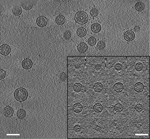 Figure 1. Cryo-electron tomography of MHV. (Bárcena M.; et al. 2009)
Cryo-ET technology ensures that the structures observed are native because it does not require additional sample treatments such as dehydration, staining, or fluorescent labeling. Cryo-ET technology exploits a transmission electron microscope (TEM) to record a series of two-dimensional (2D) projection images obtained by tilting frozen-hydrated biological samples, and then reconstructs 3D structures of the sample using computational methods. By extracting individual slices in different orientations, the morphology of the viral particles can be understood more comprehensively.
We currently do not accept samples of native coronavirus particles that could cause large-scale human infections. The samples we support for observation include coronaviruses without strong infectivity and coronavirus-like particles for various applications. Our cryo-ET service for coronavirus research has many advantages:
Scientists from Creative Biostructure can also provide you with professional technical support in experiment design, setting up data collection strategies, and image processing. If you are interested in our cryo-ET services for coronavirus-like particles, please feel free to contact us for more details. And our customer service representatives are available 24 hours a day from Monday to Sunday.
Contact us to discuss your project!
Reference
Mailing Address:
45-1 Ramsey Road, Shirley, NY 11967, USA
Email Address:
Email: info@creative-biostructure.com
Telephone Numbers:
Tel: 1-631-317-1417
Fax: 1-631-207-8356


Easy access to products and services you need from our library via powerful searching tools