Protein Structure Analysis Using Cryo-ET for Coronavirus Research
Cryo-electron tomography (Cryo-ET) is one of the three-dimensional cryogenic imaging techniques. It can help us obtain the three-dimensional (3D) structure of biological macromolecules and their complexes, organelles, cells and tissues at nanometer resolution, and it can be utilized to study the localization, distribution and interaction of macromolecules in cells. Cryo-ET has a wide range of applications in the study of virus infection. With years of experience in the analysis of the 3D structure of biological macromolecules, Creative Biostructure can use Cryo-ET technology to solve the structural analysis problems of proteins involved in your coronavirus research.
Application of Cryo-ET in Coronavirus Research
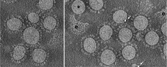
In previous studies, scientists used Cryo-ET to analyze three polymorphic coronavirus particles and virus-like particles, including Severe acute respiratory syndrome coronavirus (SARS-CoV), Mouse hepatitis virus (MHV) and Feline coronavirus (FCoV), found how membrane (M) protein (one of the structural protein of coronavirus) structure and organization is related to virus shape and size, revealing the role of M protein in assembling and determining the morphology of coronavirus. In addition, a complete image of MHV and its ribonucleoprotein was obtained by Cryo-ET.
 Figure 1. Cryo-ET images of coronavirus particles, free viral envelopes (e) and a spikeless particle (∗) are marked. (Neuman B W.; et al. 2011)
Why Do You Need This Service?
Cryogenic electron microscopy (Cryo-EM) techniques commonly used in structural virology include single particle analysis (SPA) and electron tomography (ET). SPA requires samples to be purified, homogenous and monodispersed. In contrast, Cryo-ET allows in-situ dynamic 3D structure studies because this technology is based on 3D reconstruction of two-dimensional (2D) projections from different angles of the same target without the need for averaging between particles or molecules. It is suitable for samples with more flexibility and more heterogeneity. In fact, SPA and Cryo-ET are highly complementary in structure study, and the organic combination of the two is a promising approach to study the in-situ high-resolution 3D structures of heterogeneous and flexible complexes between viruses and host factors.
What We Can Do?
For some hard-to-purify complexes involved in coronavirus studies, we recommend using Cryo-ET to obtain their structure and location information. Creative Biostructure can use cutting-edge approaches to improve resolution without damaging samples. Our service about protein structure analysis using Cryo-ET for coronavirus research includes, but is not limited to, vitrified sample preparation, focused ion beam (FIB), Cryo-ET imaging, image processing and structural analysis. To obtain structures with near-atomic resolution, we support the latest image processing method, the subtomogram averaging (STA) analysis.
Why Do You Choose Us?
- Advanced imaging equipment and image processing software
- Thick samples can be processed with FIB method
- Nanoscale information about macromolecular complexes in their natural cellular environment
- Visualization of protein-cell interactions, allowing capture of transient and dynamic processes
- Our customer service representatives are available 24 hours a day from Monday to Sunday
Contact us to discuss your project!
References
- Bárcena M.; et al. Cryo-electron tomography of mouse hepatitis virus: insights into the structure of the coronavirion. Proceedings of the National Academy of Sciences. 2009, 106(2): 582-587.
- Neuman B W.; et al. A structural analysis of M protein in coronavirus assembly and morphology. Journal of Structural Biology. 2011, 174(1): 11-22.

 Figure 1. Cryo-ET images of coronavirus particles, free viral envelopes (e) and a spikeless particle (∗) are marked. (Neuman B W.; et al. 2011)