Ultra-thin Section Electron Microscopy for Coronavirus-like Particle
The ultra-thin section electron microscopy (EM) technology is an effective means to observe the characteristics of viruses and the interaction between viruses and host cells. Creative Biostructure provides ultra-thin section EM services combined with positive staining technology to help you obtain ultrastructure information on the interaction of coronavirus-like particles with tissues or cells.
Application of Ultra-thin Section EM in Virology
Transmission electron microscopy (TEM) is an important approach for studying viruses. It is also one of the preferred technologies for the diagnosis of unknown pathogens and the identification of pathogens for bioterrorism attacks. TEM has played a crucial role in the discovery and identification of hepatitis B virus (HBV), rotavirus, SARS-CoV, and many other viruses. Negative staining and ultra-thin section are significant techniques to constitute the biological TEM technology system. Liquid samples are mainly detected by negative staining EM, while solid samples such as tissues and cultured cells are mainly detected by ultra-thin section EM. Both negative and positive staining techniques involve incubating the sample with a solution of heavy metal salts to enhance the contrast of biological samples (tissue, cell cultures, and viruses, etc.). Ultra-thin section of virus-infected/virus-like particle (VLP)-incubated tissues or cell cultures combined with a positive staining technique can provide information about the localization and the interaction of the virus/VLP in the cellular environment.
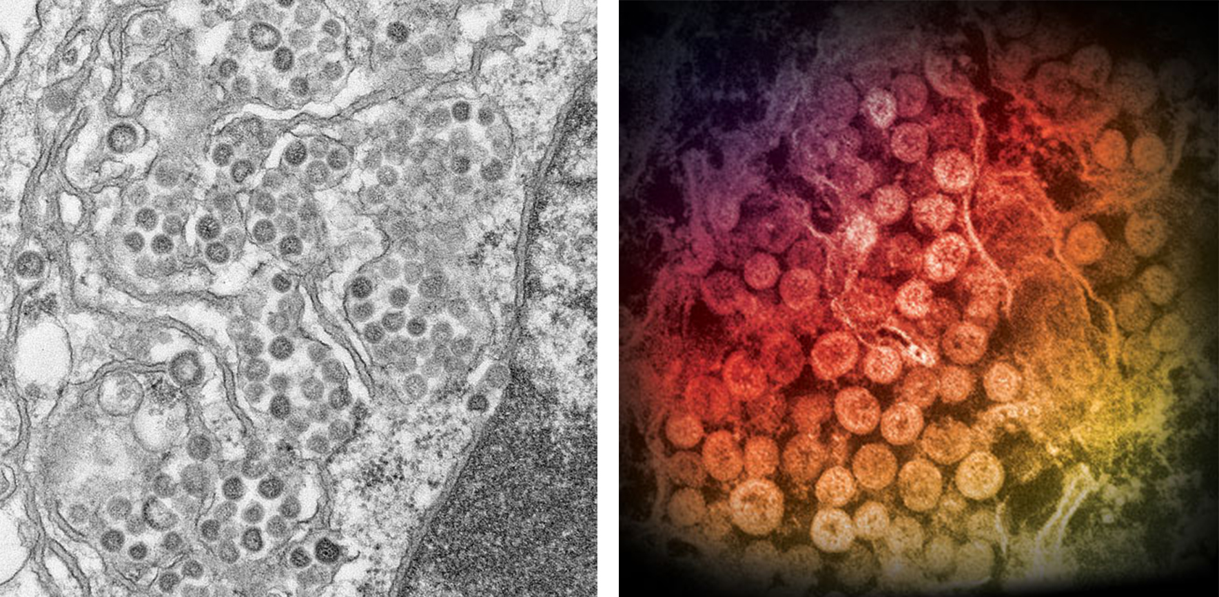
 Figure 1. Electron micrographs of ultra-thin sections of MERS-CoV. (CDC: Maureen Metcalfe/Cynthia Goldsmith/Azaibi Tamin)
Our Ultra-thin Section EM Services for Coronavirus-like Particle
Due to public safety concerns, we do not support human coronavirus samples that can be highly contagious, and our services are only available for research purposes. Our ultra-thin section EM service is suitable for the observation of coronaviruses without strong infectivity and the characterization of coronavirus-like particles used for vaccine or diagnostic reagent development. The general workflow of our ultra-thin section EM is as follows:
- The tissue or cell sample is successively fixed in glutaraldehyde and osmium tetroxide
- The fixed sample is dehydrated in graded ethanol
- Embedded flat in epoxy resins
- Ultra-thin sections are cut parallel to the surface of the dish with a diamond knife
- The grid containing the ultra-thin section of the sample is incubated in uranyl acetate and lead citrate, and then washed and dried
- Observed and photographed by TEM
Scientists from Creative Biostructure can design optimal sample preparation and data collection solutions according to your experimental samples and research needs. If you are interested in our ultra-thin section EM services for coronavirus-like particles, please feel free to contact us. And our customer service representatives are available 24 hours a day from Monday to Sunday.
Contact us to discuss your project!
Reference
- Barreto-Vieira D F, Barth O M. Negative and positive staining in transmission electron microscopy for virus diagnosis. InTech, 2015.

 Figure 1. Electron micrographs of ultra-thin sections of MERS-CoV. (CDC: Maureen Metcalfe/Cynthia Goldsmith/Azaibi Tamin)