Genetically Engineered Exosome Surface Display Service
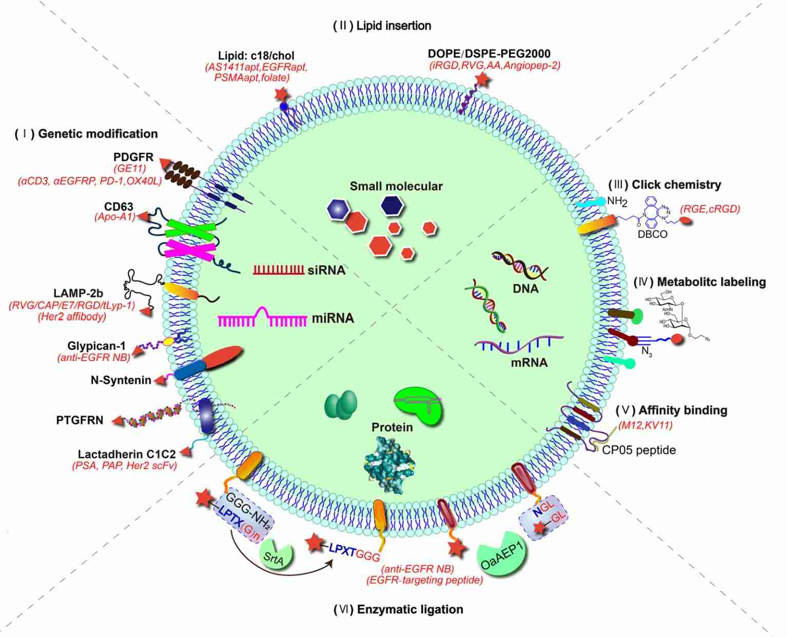
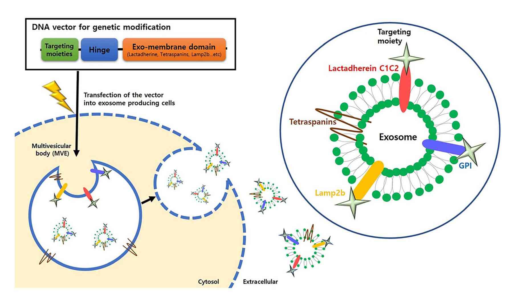
Genetic engineering represents one of the most robust and versatile approaches for exosome surface modification. By introducing engineered gene constructs into exosome-producing cells, this method enables the stable expression and incorporation of targeting ligands onto the exosomal membrane during vesicle biogenesis, resulting in homogeneous display and consistent functional performance.
At Creative Biostructure, we provide comprehensive genetically engineered exosome surface display services utilizing validated scaffold proteins including LAMP2B, CD63, CD81, CD9, PTGFRN, and proprietary platforms. Our solutions support diverse research applications ranging from targeted delivery studies and immunotherapy development to biomarker discovery and vaccine design.
Why Choose Genetic Engineering for Exosome Surface Display
While exosomes possess intrinsic biocompatibility and natural delivery capabilities, their native membrane composition often lacks sufficient specificity for precise targeting applications. Genetic engineering provides a powerful solution by enabling controlled and consistent surface modification that overcomes the limitations of native vesicles.
Key Advantages of Genetic Engineering Approach
- Stable and homogeneous display of targeting ligands across vesicle populations
- Endogenous incorporation during exosome biogenesis, preserving vesicle integrity
- High copy number display of proteins on exosome surface (up to hundreds of copies per vesicle)
- Multivalent binding capability significantly enhances target avidity and cellular uptake
- Excellent batch-to-batch consistency through stable cell line development
- Scalable production suitable for both research and preclinical applications
- Freedom from post-isolation chemical reactions that may damage vesicle structure
 Figure 1. Genetically Engineered Exosomes for miRNA Delivery to Chondrocytes in Osteoarthritis Treatment. (Liang Y, et al., 2021)
Genetic Engineering vs. Chemical Modification
| Feature | Genetic Engineering | Chemical Modification |
|---|---|---|
| Display Consistency | High and homogeneous | Variable, batch-dependent |
| Vesicle Integrity | Fully preserved | May be compromised |
| Ligand Orientation | Well-controlled, N-terminal display | Random, less defined |
| Production Scale | Easily scalable | Limited by reaction scale |
| Applicable Ligands | Proteins, peptides, antibodies | Wide range (peptides, aptamers, small molecules) |
Our Genetic Engineering Platforms
We offer multiple validated scaffold platforms for exosome surface display, each with distinct characteristics suited for different applications. Our team will recommend the optimal scaffold based on your specific target, ligand type, and experimental requirements.
Scaffold Protein Options
| Scaffold Protein | Description & Applications | Display Efficiency |
|---|---|---|
| LAMP2B | Most widely used scaffold; N-terminal fusion for extracellular domain display. Ideal for peptide ligands, targeting moieties, and antibody fragments. Extensively validated in CNS targeting and tumor models. | High |
| CD63 | Tetraspanin protein with extracellular loops for ligand fusion. Suitable for membrane proteins and protein domains. Commonly used for biosensor development and molecular interaction studies. | Moderate-High |
| CD9 | Highly abundant tetraspanin in exosomes. Excellent for protein display and functional studies. | High |
| CD81 | Tetraspanin scaffold with excellent membrane integration. Suitable for various protein fusions including immune modulators and targeting domains. | High |
| PTGFRN | Prostaglandin F2 receptor negative regulator. Demonstrates excellent protein display capacity. Validated for multi-specific therapeutic protein display with high copy numbers. | Very High |
| VSVG | Vesicular stomatitis virus glycoprotein. Viral-derived scaffold with excellent membrane incorporation. Superior for improved protein display and enhanced target cell uptake. | Very High |
| Lactadherin (C1C2) | Milk fat globule E8 domain. Good for phosphatidylserine-binding and protein domain display. Useful for targeting studies and vaccine development. | Moderate |
| LEAP Scaffold | Late domain-based exosomal antibody surface display platform. Enables bispecific antibody display for T-cell engagement and immune therapy applications. | Very High |
Displayable Ligand Types
Our genetic engineering platforms support a wide range of targeting ligands:
| Ligand Category | Examples |
|---|---|
| Targeting Peptides | RVG (CNS targeting), iRGD (tumor penetration), TLyP-1 (lung cancer), chondrocyte affinity peptide, MSC affinity peptide |
| Antibody Fragments | scFv, Fab, bispecific antibodies, immune checkpoint inhibitors (PD-L1, CD3, CTLA-4) |
| Cytokines & Growth Factors | IL-3 (CML targeting), IL-2, EGF, VEGF fragments, interferon variants |
| Receptor Domains | Single-chain receptor constructs, ligand-binding domains, SIRPα variants |
| Fluorescent Proteins | GFP, mCherry, luciferase variants for tracking and imaging studies |
Service Workflow
- Project Consultation & Target Analysis: Evaluation of target receptors, ligand sequences, cell source selection, and application goals
- Scaffold Selection & Vector Design: Optimization of scaffold protein, linker design, and fusion construct architecture
- Cell Line Engineering: Generation of stable or transient expression cell lines using validated transfection protocols
- Exosome Production & Isolation: Scalable production and purification using ultracentrifugation, SEC, or immunoaffinity methods
- Surface Display Validation: Confirmation of ligand expression and surface localization via Western blot, flow cytometry, and ELISA
- Comprehensive Characterization: Quality control including size distribution, morphology, and functional assays
- Data Delivery & Technical Support: Detailed technical reports and post-project consultation
 Figure 2. Genetically Engineered Exosome Surface Display Service Workflow. (Creative Biostructure)
Characterization and Quality Control
We perform rigorous quality assessment to ensure consistency, functionality, and research-grade quality:
Physicochemical Characterization
- Particle size distribution and concentration (NTA, DLS)
- Morphology analysis (TEM or Cryo-EM)
- Zeta potential measurement
- Exosome marker validation (CD9, CD63, CD81)
Surface Display Verification
- Western blot analysis of fusion protein expression
- Flow cytometry analysis of exosome surface
- ELISA for ligand density quantification
- Immuno-TEM for surface localization confirmation
Functional Validation
- Target binding affinity analysis
- Cellular uptake studies in target cell models
- Competitive binding assays
- In vitro and in vivo functional studies (optional)
Applications
Genetically engineered exosomes with surface display capabilities support diverse research applications:
| Application Area | Description |
|---|---|
| Targeted Drug Delivery |
|
| Immunotherapy Development |
|
| Vaccine Development |
|
| Molecular Biology & Diagnostics |
|
How to Start Your Project
We offer flexible project entry options to support different research scenarios:
| Option | Description |
|---|---|
| Client-Provided Ligand | We perform vector construction and exosome engineering using your provided ligand sequence or protein. |
| Full-Service Workflow | Complete end-to-end service including ligand identification, scaffold selection, vector design, cell engineering, exosome production, and validation. |
| Cell Source Option | Use your existing cell line or select from our validated exosome-producing cell panels (HEK293, MSC, DC, etc.). |
Project Kickoff Information
| Information Type | Details Needed |
|---|---|
| Target Information | Target cell type, receptor, tissue/organ of interest |
| Ligand Details | Targeting ligand sequence (if available), or targeting concept |
| Application Goal | Delivery study, immunotherapy, vaccine, diagnostics, etc. |
| Scale Requirements | Research-scale or preclinical-scale production needs |
What Deliverables Will You Receive
| Deliverable | Description |
|---|---|
| Engineered Exosomes | Surface-display exosomes with defined concentration, volume, and storage conditions |
| Characterization Report | Size distribution, morphology, surface marker validation, and ligand display confirmation |
| Vector Construct | Plasmid DNA encoding fusion protein (optional delivery format) |
| Cell Line | Stably engineered producer cell line (optional delivery format) |
| Functional Data | Binding and uptake performance data, optional functional validation results |
| Technical Report | Detailed methodology, optimization parameters, and data interpretation |
Why Choose Creative Biostructure
- Multiple validated scaffold platforms, including LAMP2B, CD9, CD63, CD81, PTGFRN, VSVG, and proprietary systems
- Advanced genetic engineering expertise for stable cell line development and scalable production
- Comprehensive characterization suite: NTA, DLS, TEM, Western blot, flow cytometry, ELISA, and functional assays
- High batch-to-batch consistency through rigorous QC protocols and standardized workflows
- Flexible project design supporting early-stage research to preclinical development
- Integration capability with cargo loading, PEGylation, and other exosome modification services
- Experienced scientific team with CRO project support and post-project consultation
Case Study
Case: GEMINI-Exosomes for Targeted Cancer Immunotherapy
Introduction
Researchers developed GEMINI-Exosomes, engineered exosomes featuring surface-displayed monoclonal antibodies targeting T-cell CD3 and EGFR, alongside immune checkpoint modulators PD-1 and OX40L. These exosomes were genetically engineered to activate T-cells against EGFR-positive triple-negative breast cancer (TNBC) cells.
Results
- In vitro: GEMINI-Exosomes significantly increased the activation of CD8+ T cells and IL-2 secretion in response to EGFR-positive TNBC cells.
- In vivo: In a mouse model, GEMINI-Exosomes induced potent tumor growth inhibition in TNBC, demonstrating strong anti-tumor immunity without significant systemic toxicity.
- Targeted Activity: The exosomes exhibited enhanced targeting of PD-L1 and OX40 pathways, modulating immune responses in the tumor microenvironment.
 Figure 3. Immunoblot, size distribution, ELISA, flow cytometry, and T-cell activation assays characterizing aCD3-aEGFR-PD-1-OX40L GEMINI-Exosomes, showing binding to PD-L1, PD-L2, and OX40, and enhanced IL-2 secretion. (Cheng Q, et al., 2022)
Conclusion
GEMINI-Exosomes are a promising platform for cancer immunotherapy, offering a genetically engineered, multifunctional exosome solution for activating and modulating T-cell immunity against specific tumor antigens. The study highlights their potential for enhancing therapeutic efficacy in cancer treatment.
Ready to develop genetically engineered exosomes for your research? Our expert team will design a tailored solution based on your specific targets and experimental goals. Contact us to discuss your project and accelerate your exosome engineering workflow.
References
- Liang Y, Duan L, Lu J, et al. Engineering exosomes for targeted drug delivery. Theranostics. 2021, 11(7): 3183.
- Cheng Q, Dai Z, Smbatyan G, et al. Eliciting anti-cancer immunity by genetically engineered multifunctional exosomes. Molecular Therapy. 2022, 30(9): 3066-3077.
- Chen R, Kang Z, Li W, et al. Extracellular vesicle surface display of αPD‐L1 and αCD3 antibodies via engineered late domain‐based scaffold to activate T‐cell anti‐tumor immunity. Journal of Extracellular Vesicles. 2024, 13(7): e12490.
Frequently Asked Questions
For any inquiries, our support team is ready to help you get technical support for your research and maximize your experience with Creative Biostructure.