Exosome Characterization Services
Exosomes are first found in the supernatant of sheep red blood cells cultured in vitro. They are vesicles secreted by cells with relatively uniform size, a diameter of 30~160 nanometers and a density of 1.10~1.18 g/mL. Exosome carries a variety of proteins, mRNAs and miRNAs, and participate in the processes of cell communication, cell migration, angiogenesis and tumor cell growth, and may become a natural carrier of drugs for clinical treatment. Isolation and characterization of exosomes are technically challenging due to the persistence of contaminants such as microcapsules, lipoprotein particles, microorganisms, microbodies, protein aggregates, and DNA and other necrosis products during purification. Therefore, exosome characterization techniques are of critical importance in the exosome field.
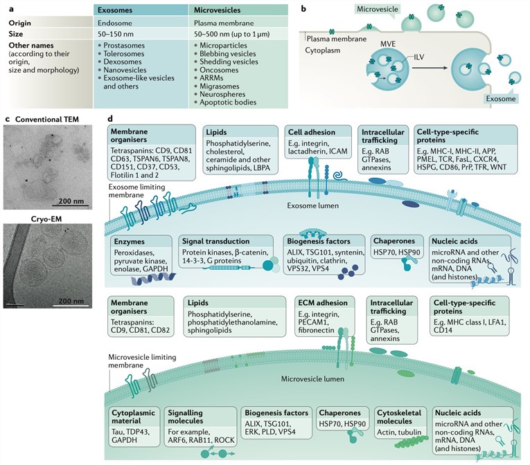 Figure 1. Main features of extracellular vesicles. (Van Niel, et al. 2018)
Creative Biostructure provides exosome characterization services for our customers. Our exosome research team has many years of experience in exosome research and can provide you with a range of services including exosome isolation, purification, identification, characterization and so on.
Various methods for exosome characterization:
- Visualization by Transmission Electron Microscopy
- Nanoparticle Tracking Analysis
- Detection of Marker Proteins by Western Blot
- Detection of Marker Proteins by Flow Cytometry
- MRI-based Exosome Quantification
Transmission electron microscope (TEM) is a kind of electronic optical instrument with high resolution and high magnification, which uses the electron beam with a very short wavelength as the illumination source and focuses on imaging with the electromagnetic lens. Electron microscope imaging can directly see the morphology of particles, so it can be used to identify the existence and integrity of exosomes.
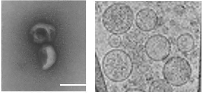 Figure 2. Electron micrographs of exosomes. Left: TEM imaging; Right: Cryo-TEM imaging. (Chen Zhang, et al. 2020; Anton Emelyanov, et al. 2020)
Nanoparticle tracking analysis (NTA) is a novel method to calculate the particle size distribution and concentration. It senses the presence of particles through optical principle, so it can confirm the population characteristics of the number and diameter of "exosomes" in samples. However, it is impossible to confirm whether the detected particles are exosomes, reflect the particle morphology, and distinguish exosomes from other nanoparticles.
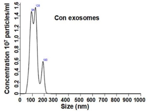 Figure 3. Nanoparticle tracking analysis. (Chen Zhang, et al. 2020)
Exosomes were identified by WB assay to detect the expression of biomarker proteins such as TSG101, CD81, CD63 and CD9 proteins or some other specific protein on exosomes.
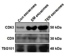 Figure 4. Western blot of marker proteins CD9, CD63, CD81, and TSG101. (Chen Zhang, et al. 2020)
In addition to WB, marker proteins in exosomes can also be detected by flow cytometry.
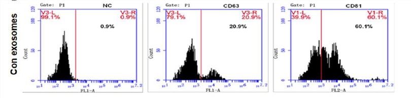 Figure 5. Flow cytometry of marker proteins CD9, CD63, CD81, and TSG101. (Chen Zhang, et al. 2020)
Recent studies have shown that novel methods of labeling exosomes with nanomaterials can be traceable by magnetic resonance in vivo. Currently, common molecular imaging methods for exosome tracers include optical imaging, magnetic resonance imaging, radionuclide molecular imaging, photoacoustic imaging, and multimodal imaging, etc. The development of these imaging methods is of great significance for the study of the role of exosomes in clinical diagnosis and treatment, especially in the integration of tumor diagnosis and treatment.
Our service content:

From sampling protocols, database building and sequencing, to analysis strategies, Creative Biostructure can tailor solutions to your specific research purpose.
If you are interested in our exosome characterization services, please feel free to contact us. We are looking forward to cooperating with you.
Ordering Process

References
- Van Niel, et al. Shedding light on the cell biology of extracellular vesicles. Nature Reviews Molecular Cell Biology. 2018, 19(4): 213-228.
- Chen Zhang, et al. Plasma exosomal miR-375-3p regulates mitochondria-dependent keratinocyte apoptosis by targeting XIAP in severe drug-induced skin reactions. Science Translational Medicine. Sci Transl Med. 2020, 12(574): eaaw6142.
- Anton Emelyanov, et al. Cryo-electron microscopy of extracellular vesicles from cerebrospinal fluid. PLoS One. 2020, 15(1): e0227949.

