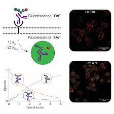
Selecting the most effective molecules for drug delivery usually requires trial and error. Recently, researchers at Cornell University have revealed how to transport molecules behave in living cells, providing some basis for judgment.
The drug delivery system controls when and where the drug is released in the body. The principle of many drug delivery systems is to connect antibodies (looking for targets such as cancer cells) with drugs used to destroy that target. On the one hand, it ensures that the drug is bound to the antibody before entering the target cell; on the other hand, it must be released into the cell in the right place at the right time.

By testing in a cell-free environment, researchers were able to gather clues about the effectiveness of such “connections”. It is easier to study because it does not include complex interactions in living cells, but the accuracy is relatively low, which may lead to failure in subsequent tests.
In this regard, a research group led by Chris Alabi, Associate Professor of Chemical and Biomolecular Engineering, developed a method that uses fluorescent probes to observe and measure the rate at which “adapters” successfully release drugs in living cells. The results were published recently in the journal Cell Chemical Biology.
“With our technology, pharmaceutical companies can make informed decisions based on actual intracellular responses before integrating drug systems together,” said Alabi.
This probe consists of two fluorescent dyes that are not visible to each other when they are bundled with antibodies as part of a transmission system. When the adapters broke and decolorized the dye, they became visible under the microscope, indicating that the drug had been released intracellularly.
These probes were developed in the lab of Alabi and they provide engineers with a precise way to measure the rate at which chemical bonds in the adapters break from the delivery system while inside the cell.
To demonstrate the probe, Alabi and his team designed an antibody-anchored molecule with a disulfide bond that is known to work well in the HER2 protein, a very valuable target in breast cancer therapy. Once the delivery system reached the protein, the team was able to reliably measure intracellular processes such as the kinetics and half-life of disulfide bonds.
Reference
Michelle R. Sorkin, Joshua A. Walker, Sneha R. Kabaria, Nicole P. Torosian, Christopher A. Alabi. Responsive Antibody Conjugates Enable Quantitative Determination of Intracellular Bond Degradation Rate. Cell Chemical Biology, 2019; DOI: 10.1016/j.chembiol.2019.09.008